[English] 日本語
Yorodumi
- PDB-6yxa: Structure of the bifunctional Rel enzyme from B. subtilis -

+
Open data


ID or keywords:

Loading...

-
Basic information

Entry
Database: PDB / ID: 6yxa
TitleStructure of the bifunctional Rel enzyme from B. subtilis
ComponentsGTP pyrophosphokinase
KeywordsSIGNALING PROTEIN / bifunctional (p)ppGpp synthease/hydrolase / stringent response / ribosome interacting
Function / homology
Function and homology information


GTP diphosphokinase activity / guanosine tetraphosphate biosynthetic process / GTP diphosphokinase / guanosine-3',5'-bis(diphosphate) 3'-diphosphatase activity / response to starvation / kinase activity / phosphorylation / GTP binding / ATP binding / plasma membrane
Similarity search - Function
RelA/SpoT, AH and RIS domains / RelA/SpoT, AH and RIS domains / HD domain / RelA/SpoT family / RelA/SpoT, TGS domain / ACT domain / Region found in RelA / SpoT proteins / RelA/SpoT / Region found in RelA / SpoT proteins / TGS domain ...RelA/SpoT, AH and RIS domains / RelA/SpoT, AH and RIS domains / HD domain / RelA/SpoT family / RelA/SpoT, TGS domain / ACT domain / Region found in RelA / SpoT proteins / RelA/SpoT / Region found in RelA / SpoT proteins / TGS domain / ACT domain profile. / ACT domain / ACT-like domain / HD domain profile. / HD domain / TGS domain profile. / TGS / TGS-like / HD domain / Beta-grasp domain superfamily / Metal dependent phosphohydrolases with conserved 'HD' motif. / HD/PDEase domain / Nucleotidyltransferase superfamily
Similarity search - Domain/homology
: / GTP pyrophosphokinase
Similarity search - Component
Biological speciesBacillus subtilis subsp. subtilis str. 168 (bacteria)
MethodX-RAY DIFFRACTION / SYNCHROTRON / MOLECULAR REPLACEMENT / Resolution: 3.95 Å
AuthorsPausch, P. / Bange, G.
Funding support Germany, 1items
OrganizationGrant numberCountry
German Research Foundation (DFG)SPP1879 Germany
Citation
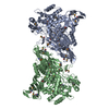
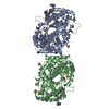
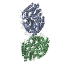
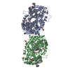
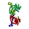
Journal: Cell Rep / Year: 2020
Title: Structural Basis for Regulation of the Opposing (p)ppGpp Synthetase and Hydrolase within the Stringent Response Orchestrator Rel.
Authors: Patrick Pausch / Maha Abdelshahid / Wieland Steinchen / Heinrich Schäfer / Fabio Lino Gratani / Sven-Andreas Freibert / Christiane Wolz / Kürşad Turgay / Daniel N Wilson / Gert Bange /
Abstract: The stringent response enables metabolic adaptation of bacteria under stress conditions and is governed by RelA/SpoT Homolog (RSH)-type enzymes. Long RSH-type enzymes encompass an N-terminal domain ...The stringent response enables metabolic adaptation of bacteria under stress conditions and is governed by RelA/SpoT Homolog (RSH)-type enzymes. Long RSH-type enzymes encompass an N-terminal domain (NTD) harboring the second messenger nucleotide (p)ppGpp hydrolase and synthetase activity and a stress-perceiving and regulatory C-terminal domain (CTD). CTD-mediated binding of Rel to stalled ribosomes boosts (p)ppGpp synthesis. However, how the opposing activities of the NTD are controlled in the absence of stress was poorly understood. Here, we demonstrate on the RSH-type protein Rel that the critical regulative elements reside within the TGS (ThrRS, GTPase, and SpoT) subdomain of the CTD, which associates to and represses the synthetase to concomitantly allow for activation of the hydrolase. Furthermore, we show that Rel forms homodimers, which appear to control the interaction with deacylated-tRNA, but not the enzymatic activity of Rel. Collectively, our study provides a detailed molecular view into the mechanism of stringent response repression in the absence of stress.
History
DepositionApr 30, 2020Deposition site: PDBE / Processing site: PDBE
Revision 1.0Sep 23, 2020Provider: repository / Type: Initial release
Revision 1.1Sep 30, 2020Group: Database references / Derived calculations
Category: citation / citation_author ...citation / citation_author / pdbx_struct_assembly / pdbx_struct_assembly_gen / pdbx_struct_assembly_prop / pdbx_struct_oper_list
Item: _citation.journal_volume / _citation.page_first ..._citation.journal_volume / _citation.page_first / _citation.page_last / _citation.pdbx_database_id_PubMed / _citation.title / _citation_author.name / _pdbx_struct_assembly.details / _pdbx_struct_assembly.method_details / _pdbx_struct_assembly.oligomeric_count / _pdbx_struct_assembly.oligomeric_details
Revision 1.2Jan 24, 2024Group: Data collection / Database references / Refinement description
Category: chem_comp_atom / chem_comp_bond ...chem_comp_atom / chem_comp_bond / database_2 / pdbx_initial_refinement_model
Item: _database_2.pdbx_DOI / _database_2.pdbx_database_accession

-
Structure visualization

Structure viewerMolecule:
MolmilJmol/JSmol

Downloads & links

-
Assembly

Deposited unit
A: GTP pyrophosphokinase
hetero molecules


Theoretical massNumber of molelcules
Total (without water)65,3352
Polymers65,2801
Non-polymers551
Water00
1
A: GTP pyrophosphokinase
hetero molecules

A: GTP pyrophosphokinase
hetero molecules


Theoretical massNumber of molelcules
Total (without water)130,6694
Polymers130,5592
Non-polymers1102
Water0
TypeNameSymmetry operationNumber
identity operation1_555x,y,z1
crystal symmetry operation7_555y,x,-z1
Unit cell
Length a, b, c (Å)130.152, 130.152, 157.621
Angle α, β, γ (deg.)90.000, 90.000, 90.000
Int Tables number96
Space group name H-MP43212
Space group name HallP4nw2abw

-
Components

#1: Protein GTP pyrophosphokinase / (p)ppGpp synthase / ATP:GTP 3'-pyrophosphotransferase / ppGpp synthase I


Mass: 65279.621 Da / Num. of mol.: 1
Source method: isolated from a genetically manipulated source
Source: (gene. exp.) Bacillus subtilis subsp. subtilis str. 168 (bacteria)
Gene: relA, BSU27600 / Production host: Escherichia coli BL21(DE3) (bacteria) / References: UniProt: O54408, GTP diphosphokinase
#2: Chemical ChemComp-MN / MANGANESE (II) ION


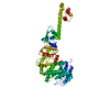
Mass: 54.938 Da / Num. of mol.: 1 / Source method: obtained synthetically / Formula: Mn
Has ligand of interestN

-
Experimental details

-
Experiment

ExperimentMethod: X-RAY DIFFRACTION / Number of used crystals: 1

-
Sample preparation

CrystalDensity Matthews: 5.37 Å3/Da / Density % sol: 77.12 %
Crystal growTemperature: 293 K / Method: vapor diffusion, sitting drop
Details: 1 M Lithium chloride, 0.1 M Bicine pH 9.0, 10% PEG6000 (w/v), final pH 9.0

-
Data collection

DiffractionMean temperature: 100 K / Serial crystal experiment: N
Diffraction sourceSource: SYNCHROTRON / Site: ESRF / Beamline: ID29 / Wavelength: 0.979 Å
DetectorType: DECTRIS PILATUS 6M / Detector: PIXEL / Date: Feb 15, 2017
RadiationProtocol: SINGLE WAVELENGTH / Monochromatic (M) / Laue (L): M / Scattering type: x-ray
Radiation wavelengthWavelength: 0.979 Å / Relative weight: 1
ReflectionResolution: 3.95→48.72 Å / Num. obs: 12337 / % possible obs: 99.28 % / Redundancy: 14.2 % / Biso Wilson estimate: 188.71 Å2 / Rpim(I) all: 0.043 / Net I/σ(I): 10.04
Reflection shellResolution: 3.95→4.09 Å / Num. unique obs: 1158 / Rpim(I) all: 0.528

-
Processing

Software
NameVersionClassification
PHENIX1.18_3845refinement
XDSdata reduction
Aimlessdata scaling
PHASERphasing
RefinementMethod to determine structure: MOLECULAR REPLACEMENT
Starting model: 1VJ7, chain B
Resolution: 3.95→48.72 Å / SU ML: 0.5149 / Cross valid method: FREE R-VALUE / σ(F): 1.34 / Phase error: 32.3858
Stereochemistry target values: GeoStd + Monomer Library + CDL v1.2
RfactorNum. reflection% reflection
Rfree0.2839 1206 9.82 %
Rwork0.2635 11077 -
obs0.2655 12283 99.15 %
Solvent computationShrinkage radii: 0.9 Å / VDW probe radii: 1.11 Å / Solvent model: FLAT BULK SOLVENT MODEL
Displacement parametersBiso mean: 224.56 Å2
Refinement stepCycle: LAST / Resolution: 3.95→48.72 Å
ProteinNucleic acidLigandSolventTotal
Num. atoms4368 0 1 0 4369
Refine LS restraints
Refine-IDTypeDev idealNumber
X-RAY DIFFRACTIONf_bond_d0.00264453
X-RAY DIFFRACTIONf_angle_d0.55186002
X-RAY DIFFRACTIONf_chiral_restr0.0394659
X-RAY DIFFRACTIONf_plane_restr0.0034773
X-RAY DIFFRACTIONf_dihedral_angle_d13.23511701
LS refinement shell
Resolution (Å)Rfactor RfreeNum. reflection RfreeRfactor RworkNum. reflection RworkRefine-ID% reflection obs (%)
3.95-4.110.3691050.35231139X-RAY DIFFRACTION92.35
4.11-4.30.3541390.31561199X-RAY DIFFRACTION100
4.3-4.520.28911310.2591207X-RAY DIFFRACTION100
4.52-4.810.28531300.25521222X-RAY DIFFRACTION99.93
4.81-5.180.28331250.2531237X-RAY DIFFRACTION100
5.18-5.70.32511320.28471249X-RAY DIFFRACTION100
5.7-6.520.36291510.30011219X-RAY DIFFRACTION100
6.52-8.210.28271550.26751245X-RAY DIFFRACTION100
8.21-48.720.23671380.2391360X-RAY DIFFRACTION99.93
Refinement TLS params.Method: refined / Origin x: -29.1278063713 Å / Origin y: -11.1200865575 Å / Origin z: -5.17262232223 Å
111213212223313233
T1.15358006448 Å2-0.211113243654 Å2-0.204871638788 Å2-1.22647303877 Å20.0554197779487 Å2--0.853318675977 Å2
L2.43561523171 °2-2.3840564213 °2-0.218483459302 °2-9.21029594531 °20.609182535281 °2--2.41946534807 °2
S0.0330908197326 Å °-0.391454701644 Å °0.142315024482 Å °0.24082870668 Å °0.142462941214 Å °-0.062531345001 Å °-0.18513106863 Å °0.113047993707 Å °-0.206019858312 Å °
Refinement TLS groupSelection details: all

+
About Yorodumi

-
News

-
Feb 9, 2022. New format data for meta-information of EMDB entries

New format data for meta-information of EMDB entries

  • Version 3 of the EMDB header file is now the official format.
  • The previous official version 1.9 will be removed from the archive.

Related info.:EMDB header

External links:wwPDB to switch to version 3 of the EMDB data model

-
Aug 12, 2020. Covid-19 info

Covid-19 info

URL: https://pdbjlvh1.pdbj.org/emnavi/covid19.php

New page: Covid-19 featured information page in EM Navigator.

Related info.:Covid-19 info / Mar 5, 2020. Novel coronavirus structure data

+
Mar 5, 2020. Novel coronavirus structure data

Novel coronavirus structure data

Related info.:Yorodumi Speices / Aug 12, 2020. Covid-19 info

External links:COVID-19 featured content - PDBj / Molecule of the Month (242):Coronavirus Proteases

+
Jan 31, 2019. EMDB accession codes are about to change! (news from PDBe EMDB page)

EMDB accession codes are about to change! (news from PDBe EMDB page)

  • The allocation of 4 digits for EMDB accession codes will soon come to an end. Whilst these codes will remain in use, new EMDB accession codes will include an additional digit and will expand incrementally as the available range of codes is exhausted. The current 4-digit format prefixed with “EMD-” (i.e. EMD-XXXX) will advance to a 5-digit format (i.e. EMD-XXXXX), and so on. It is currently estimated that the 4-digit codes will be depleted around Spring 2019, at which point the 5-digit format will come into force.
  • The EM Navigator/Yorodumi systems omit the EMD- prefix.

Related info.:Q: What is EMD? / ID/Accession-code notation in Yorodumi/EM Navigator

External links:EMDB Accession Codes are Changing Soon! / Contact to PDBj

+
Jul 12, 2017. Major update of PDB

Major update of PDB

  • wwPDB released updated PDB data conforming to the new PDBx/mmCIF dictionary.
  • This is a major update changing the version number from 4 to 5, and with Remediation, in which all the entries are updated.
  • In this update, many items about electron microscopy experimental information are reorganized (e.g. em_software).
  • Now, EM Navigator and Yorodumi are based on the updated data.

External links:wwPDB Remediation / Enriched Model Files Conforming to OneDep Data Standards Now Available in the PDB FTP Archive

-
Yorodumi

Thousand views of thousand structures

  • Yorodumi is a browser for structure data from EMDB, PDB, SASBDB, etc.
  • This page is also the successor to EM Navigator detail page, and also detail information page/front-end page for Omokage search.
  • The word "yorodu" (or yorozu) is an old Japanese word meaning "ten thousand". "mi" (miru) is to see.

Related info.:EMDB / PDB / SASBDB / Comparison of 3 databanks / Yorodumi Search / Aug 31, 2016. New EM Navigator & Yorodumi / Yorodumi Papers / Jmol/JSmol / Function and homology information / Changes in new EM Navigator and Yorodumi

Read more