 Movie
Movie Controller

[English] 日本語
 Yorodumi
- PDB-6yv6: Crystal Structure of Serine protease SplB N2K/N3Q/S154R from Stap... -
+ Open data

- Basic information
| Entry | Database: PDB / ID: 6yv6 | ||||||
|---|---|---|---|---|---|---|---|
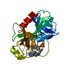
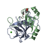
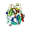
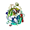
| Title | Crystal Structure of Serine protease SplB N2K/N3Q/S154R from Staphylococcus aureus | ||||||
 Components Components | Serine protease | ||||||
 Keywords Keywords | BIOSYNTHETIC PROTEIN / Spl / serine protease-like | ||||||
| Function / homology |  Function and homology information Function and homology informationHydrolases; Acting on peptide bonds (peptidases); Serine endopeptidases / serine-type endopeptidase activity / proteolysis Similarity search - Function | ||||||
| Biological species |   Staphylococcus aureus (bacteria) Staphylococcus aureus (bacteria) | ||||||
| Method |  X-RAY DIFFRACTION / SYNCHROTRON / MOLECULAR REPLACEMENT / molecular replacement / Resolution: 1.36 Å X-RAY DIFFRACTION / SYNCHROTRON / MOLECULAR REPLACEMENT / molecular replacement / Resolution: 1.36 Å | ||||||
 Authors Authors | Rangel Pereira, M.R. / Brear, P. / Knyphausen, P. / Jermutus, L. / Hollfelder, F. | ||||||
| Funding support |  United Kingdom, 1items United Kingdom, 1items
| ||||||
 Citation Citation | Journal: to be published Title: Crystal Structure of Serine protease SplB N2K/N3Q/S154R from Staphylococcus aureus Authors: Knyphausen, P. / Rangel Pereira, M.R. / Brear, P. / Jermutus, L. / Hollfelder, F. | ||||||
| History |
|
- Structure visualization
| Structure viewer | Molecule: MolmilJmol/JSmol |
|---|
- Downloads & links
-Download
| PDBx/mmCIF format | 6yv6.cif.gz | 57.5 KB | Display | PDBx/mmCIF format |
|---|---|---|---|---|
| PDB format | pdb6yv6.ent.gz | 39.8 KB | Display | PDB format |
| PDBx/mmJSON format | 6yv6.json.gz | Tree view | PDBx/mmJSON format | |
| Others |  Other downloads Other downloads |
-Validation report
| Arichive directory | https://data.pdbj.org/pub/pdb/validation_reports/yv/6yv6ftp://data.pdbj.org/pub/pdb/validation_reports/yv/6yv6 | HTTPS FTP |
|---|
-Related structure data
| Related structure data |  2vidS S: Starting model for refinement |
|---|---|
| Similar structure data |

-Links
 PDBj
PDBj- Assembly
| Deposited unit | 
| ||||||||
|---|---|---|---|---|---|---|---|---|---|
| 1 |
| ||||||||
| Unit cell |
|
-Components
| #1: Protein | Mass: 22645.434 Da / Num. of mol.: 1 / Fragment: SPLB PROTEASE / Mutation: N2K/N3Q/S154R Source method: isolated from a genetically manipulated source Source: (gene. exp.) Staphylococcus aureus (bacteria) / Gene: splB / Plasmid: pRSF / Production host: References: UniProt: A0A2D1PJH6, Hydrolases; Acting on peptide bonds (peptidases); Serine endopeptidases | ||||||
|---|---|---|---|---|---|---|---|
| #2: Chemical |   Mass: 238.278 Da / Num. of mol.: 2 / Source method: obtained synthetically / Formula: C10H22O6 / Comment: precipitant*YM Mass: 238.278 Da / Num. of mol.: 2 / Source method: obtained synthetically / Formula: C10H22O6 / Comment: precipitant*YM#3: Chemical | ChemComp-PEG / |   Mass: 106.120 Da / Num. of mol.: 1 / Source method: obtained synthetically / Formula: C4H10O3 Mass: 106.120 Da / Num. of mol.: 1 / Source method: obtained synthetically / Formula: C4H10O3#4: Water | ChemComp-HOH / |  Mass: 18.015 Da / Num. of mol.: 112 / Source method: isolated from a natural source / Formula: H2O Mass: 18.015 Da / Num. of mol.: 112 / Source method: isolated from a natural source / Formula: H2OHas ligand of interest | N | |
-Experimental details
-Experiment
| Experiment | Method: X-RAY DIFFRACTION / Number of used crystals: 1 |
|---|
- Sample preparation
| Crystal | Density Matthews: 2.03 Å3/Da / Density % sol: 39.37 % / Mosaicity: 0 ° |
|---|---|
| Crystal grow | Temperature: 298 K / Method: vapor diffusion, sitting drop / pH: 8 / Details: PEG3350 30%, TRIS-Cl pH 8.0 |
-Data collection
| Diffraction | Mean temperature: 100 K / Serial crystal experiment: N | |||||||||||||||||||||||||||
|---|---|---|---|---|---|---|---|---|---|---|---|---|---|---|---|---|---|---|---|---|---|---|---|---|---|---|---|---|
| Diffraction source | Source: SYNCHROTRON / Site: Diamond / Beamline: I04 / Wavelength: 0.9795 Å | |||||||||||||||||||||||||||
| Detector | Type: DECTRIS PILATUS 6M / Detector: PIXEL / Date: Sep 13, 2017 | |||||||||||||||||||||||||||
| Radiation | Protocol: SINGLE WAVELENGTH / Monochromatic (M) / Laue (L): M / Scattering type: x-ray | |||||||||||||||||||||||||||
| Radiation wavelength | Wavelength: 0.9795 Å / Relative weight: 1 | |||||||||||||||||||||||||||
| Reflection | Resolution: 1.36→32.63 Å / Num. obs: 37738 / % possible obs: 99.4 % / Redundancy: 7.2 % / Biso Wilson estimate: 20.31 Å2 / CC1/2: 1 / Rmerge(I) obs: 0.052 / Rpim(I) all: 0.021 / Rrim(I) all: 0.057 / Net I/σ(I): 15.2 / Num. measured all: 271636 / Scaling rejects: 18 | |||||||||||||||||||||||||||
| Reflection shell | Diffraction-ID: 1 / Redundancy: 6.6 %
|
-Phasing
| Phasing | Method: molecular replacement |
|---|
- Processing
| Software |
| ||||||||||||||||||||||||||||||||||||||||||||||||||||||||||||||||||||||||||||||||||||||||||||||||||||||||||||
|---|---|---|---|---|---|---|---|---|---|---|---|---|---|---|---|---|---|---|---|---|---|---|---|---|---|---|---|---|---|---|---|---|---|---|---|---|---|---|---|---|---|---|---|---|---|---|---|---|---|---|---|---|---|---|---|---|---|---|---|---|---|---|---|---|---|---|---|---|---|---|---|---|---|---|---|---|---|---|---|---|---|---|---|---|---|---|---|---|---|---|---|---|---|---|---|---|---|---|---|---|---|---|---|---|---|---|---|---|---|
| Refinement | Method to determine structure: MOLECULAR REPLACEMENT Starting model: 2VID Resolution: 1.36→19.36 Å / Cor.coef. Fo:Fc: 0.959 / Cor.coef. Fo:Fc free: 0.959 / SU R Cruickshank DPI: 0.061 / Cross valid method: THROUGHOUT / σ(F): 0 / SU R Blow DPI: 0.061 / SU Rfree Blow DPI: 0.059 / SU Rfree Cruickshank DPI: 0.06
| ||||||||||||||||||||||||||||||||||||||||||||||||||||||||||||||||||||||||||||||||||||||||||||||||||||||||||||
| Displacement parameters | Biso max: 110.34 Å2 / Biso mean: 28.11 Å2 / Biso min: 13.03 Å2
| ||||||||||||||||||||||||||||||||||||||||||||||||||||||||||||||||||||||||||||||||||||||||||||||||||||||||||||
| Refinement step | Cycle: final / Resolution: 1.36→19.36 Å
| ||||||||||||||||||||||||||||||||||||||||||||||||||||||||||||||||||||||||||||||||||||||||||||||||||||||||||||
| Refine LS restraints |
| ||||||||||||||||||||||||||||||||||||||||||||||||||||||||||||||||||||||||||||||||||||||||||||||||||||||||||||
| LS refinement shell | Resolution: 1.36→1.4 Å / Rfactor Rfree error: 0 / Total num. of bins used: 19
|
