 Movie
Movie Controller

[English] 日本語
 Yorodumi
- PDB-6yp6: Structural annotation of the conserved carbohydrate esterase vb_2... -
+ Open data

- Basic information
| Entry | Database: PDB / ID: 6yp6 | ||||||
|---|---|---|---|---|---|---|---|
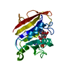
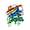
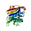
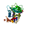
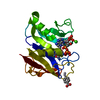
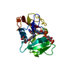
| Title | Structural annotation of the conserved carbohydrate esterase vb_24B_21 from Shiga toxin-encoding bacteriophage phi24B | ||||||
 Components Components | NanS-p | ||||||
 Keywords Keywords | SUGAR BINDING PROTEIN / Carbohydrate binding protein | ||||||
| Function / homology |  Function and homology information Function and homology information | ||||||
| Biological species |  Escherichia phage vB_EcoP_24B (virus) Escherichia phage vB_EcoP_24B (virus) | ||||||
| Method |  X-RAY DIFFRACTION / SYNCHROTRON / SAD / Resolution: 0.97 Å X-RAY DIFFRACTION / SYNCHROTRON / SAD / Resolution: 0.97 Å | ||||||
 Authors Authors | Mayans, O. / Franke, B. | ||||||
 Citation Citation | Journal: J.Struct.Biol. / Year: 2020 Title: Structural annotation of the conserved carbohydrate esterase vb_24B_21 from Shiga toxin-encoding bacteriophage Phi 24 B . Authors: Franke, B. / Veses-Garcia, M. / Diederichs, K. / Allison, H. / Rigden, D.J. / Mayans, O. | ||||||
| History |
|
- Structure visualization
| Structure viewer | Molecule: MolmilJmol/JSmol |
|---|
- Downloads & links
-Download
| PDBx/mmCIF format | 6yp6.cif.gz | 179.5 KB | Display | PDBx/mmCIF format |
|---|---|---|---|---|
| PDB format | pdb6yp6.ent.gz | 119.1 KB | Display | PDB format |
| PDBx/mmJSON format | 6yp6.json.gz | Tree view | PDBx/mmJSON format | |
| Others |  Other downloads Other downloads |
-Validation report
| Summary document | 6yp6_validation.pdf.gz | 429.5 KB | Display | wwPDB validaton report |
|---|---|---|---|---|
| Full document | 6yp6_full_validation.pdf.gz | 431 KB | Display | |
| Data in XML | 6yp6_validation.xml.gz | 15 KB | Display | |
| Data in CIF | 6yp6_validation.cif.gz | 23.4 KB | Display | |
| Arichive directory | https://data.pdbj.org/pub/pdb/validation_reports/yp/6yp6ftp://data.pdbj.org/pub/pdb/validation_reports/yp/6yp6 | HTTPS FTP |
-Related structure data
| Similar structure data |
|---|


-Links
 PDBj
PDBj- Assembly
| Deposited unit | 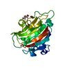
| ||||||||||||
|---|---|---|---|---|---|---|---|---|---|---|---|---|---|
| 1 |
| ||||||||||||
| Unit cell |
|
-Components
| #1: Protein | Mass: 22901.604 Da / Num. of mol.: 1 Source method: isolated from a genetically manipulated source Source: (gene. exp.) Escherichia phage vB_EcoP_24B (virus) / Gene: vb_24B_21 / Production host:  |
|---|---|
| #2: Chemical | ChemComp-GOL /   Mass: 92.094 Da / Num. of mol.: 1 / Source method: obtained synthetically / Formula: C3H8O3 Mass: 92.094 Da / Num. of mol.: 1 / Source method: obtained synthetically / Formula: C3H8O3 |
| #3: Water | ChemComp-HOH /  Mass: 18.015 Da / Num. of mol.: 431 / Source method: isolated from a natural source / Formula: H2O Mass: 18.015 Da / Num. of mol.: 431 / Source method: isolated from a natural source / Formula: H2O |
| Has ligand of interest | N |
-Experimental details
-Experiment
| Experiment | Method: X-RAY DIFFRACTION / Number of used crystals: 1 |
|---|
- Sample preparation
| Crystal | Density Matthews: 2.37 Å3/Da / Density % sol: 48.2 % |
|---|---|
| Crystal grow | Temperature: 293 K / Method: vapor diffusion, hanging drop Details: 0.9 M LiSO4, 10% [v/v] glycerol, 0.1 M Hepes pH 7.0 |
-Data collection
| Diffraction | Mean temperature: 100 K / Serial crystal experiment: N |
|---|---|
| Diffraction source | Source: SYNCHROTRON / Site: Diamond  / Beamline: I04 / Wavelength: 0.7514 Å / Beamline: I04 / Wavelength: 0.7514 Å |
| Detector | Type: ADSC QUANTUM 315 / Detector: CCD / Date: Apr 10, 2012 |
| Radiation | Protocol: SINGLE WAVELENGTH / Monochromatic (M) / Laue (L): M / Scattering type: x-ray |
| Radiation wavelength | Wavelength: 0.7514 Å / Relative weight: 1 |
| Reflection | Resolution: 0.97→25 Å / Num. obs: 128671 / % possible obs: 99.5 % / Redundancy: 7.1 % / CC1/2: 0.99 / Rsym value: 0.079 / Net I/σ(I): 14.64 |
| Reflection shell | Resolution: 0.97→0.98 Å / Mean I/σ(I) obs: 1 / Num. unique obs: 3878 / CC1/2: 0.4 / Rsym value: 1.55 |
- Processing
| Software |
| |||||||||||||||||||||||||||||||||||||||||||||||||||||||||||||||||||||||||||||||||||||||||||||||||||||||||||||||||||||||||||||||||||||||||||||||||||||||||||||||||||||||||||||||||||||||||||||||||||||||||||||||||||||||||
|---|---|---|---|---|---|---|---|---|---|---|---|---|---|---|---|---|---|---|---|---|---|---|---|---|---|---|---|---|---|---|---|---|---|---|---|---|---|---|---|---|---|---|---|---|---|---|---|---|---|---|---|---|---|---|---|---|---|---|---|---|---|---|---|---|---|---|---|---|---|---|---|---|---|---|---|---|---|---|---|---|---|---|---|---|---|---|---|---|---|---|---|---|---|---|---|---|---|---|---|---|---|---|---|---|---|---|---|---|---|---|---|---|---|---|---|---|---|---|---|---|---|---|---|---|---|---|---|---|---|---|---|---|---|---|---|---|---|---|---|---|---|---|---|---|---|---|---|---|---|---|---|---|---|---|---|---|---|---|---|---|---|---|---|---|---|---|---|---|---|---|---|---|---|---|---|---|---|---|---|---|---|---|---|---|---|---|---|---|---|---|---|---|---|---|---|---|---|---|---|---|---|---|---|---|---|---|---|---|---|---|---|---|---|---|---|---|---|---|
| Refinement | Method to determine structure: SAD / Resolution: 0.97→24.49 Å / SU ML: 0.0824 / Cross valid method: FREE R-VALUE / σ(F): 1.35 / Phase error: 15.737
| |||||||||||||||||||||||||||||||||||||||||||||||||||||||||||||||||||||||||||||||||||||||||||||||||||||||||||||||||||||||||||||||||||||||||||||||||||||||||||||||||||||||||||||||||||||||||||||||||||||||||||||||||||||||||
| Solvent computation | Shrinkage radii: 0.9 Å / VDW probe radii: 1.11 Å | |||||||||||||||||||||||||||||||||||||||||||||||||||||||||||||||||||||||||||||||||||||||||||||||||||||||||||||||||||||||||||||||||||||||||||||||||||||||||||||||||||||||||||||||||||||||||||||||||||||||||||||||||||||||||
| Displacement parameters | Biso mean: 15.71 Å2 | |||||||||||||||||||||||||||||||||||||||||||||||||||||||||||||||||||||||||||||||||||||||||||||||||||||||||||||||||||||||||||||||||||||||||||||||||||||||||||||||||||||||||||||||||||||||||||||||||||||||||||||||||||||||||
| Refinement step | Cycle: LAST / Resolution: 0.97→24.49 Å
| |||||||||||||||||||||||||||||||||||||||||||||||||||||||||||||||||||||||||||||||||||||||||||||||||||||||||||||||||||||||||||||||||||||||||||||||||||||||||||||||||||||||||||||||||||||||||||||||||||||||||||||||||||||||||
| Refine LS restraints |
| |||||||||||||||||||||||||||||||||||||||||||||||||||||||||||||||||||||||||||||||||||||||||||||||||||||||||||||||||||||||||||||||||||||||||||||||||||||||||||||||||||||||||||||||||||||||||||||||||||||||||||||||||||||||||
| LS refinement shell |
|
