 Movie
Movie Controller

[English] 日本語
 Yorodumi
- PDB-6yl2: Structural and DNA binding studies of the transcriptional repress... -
+ Open data

- Basic information
| Entry | Database: PDB / ID: 6yl2 | ||||||
|---|---|---|---|---|---|---|---|
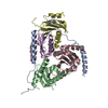
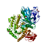
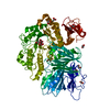
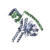
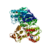
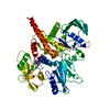
| Title | Structural and DNA binding studies of the transcriptional repressor Rv2506 (BkaR) from Mycobacterium tuberculosis supports a role in L-Leucine catabolism | ||||||
 Components Components |
| ||||||
 Keywords Keywords | DNA BINDING PROTEIN / Tuberculosis / Branch-chain amino acid catabolism / TetR Family | ||||||
| Function / homology |  Function and homology information Function and homology informationsymbiont-mediated evasion of host immune response / transcription cis-regulatory region binding / DNA-binding transcription factor activity / negative regulation of DNA-templated transcription / regulation of DNA-templated transcription / identical protein binding Similarity search - Function | ||||||
| Biological species |   Mycobacterium tuberculosis (bacteria)Mycobacterium tuberculosis H37Rv (bacteria) Mycobacterium tuberculosis (bacteria)Mycobacterium tuberculosis H37Rv (bacteria) | ||||||
| Method |  X-RAY DIFFRACTION / SYNCHROTRON / MOLECULAR REPLACEMENT / Resolution: 3.15 Å X-RAY DIFFRACTION / SYNCHROTRON / MOLECULAR REPLACEMENT / Resolution: 3.15 Å | ||||||
 Authors Authors | Keep, N.H. / Pritchard, J.E. / Sula, A. / Cole, A.R. / Kendall, S.L. | ||||||
 Citation Citation | Journal: To be published Title: Structural and DNA binding studies of the transcriptional repressor Rv2506 (BkaR) from Mycobacterium tuberculosis supports a role in L-Leucine catabolism Authors: Pritchard, J.E. / Sula, A. / Cole, A.R. / Kendall, S.L. / Keep, N.H. | ||||||
| History |
|
- Structure visualization
| Structure viewer | Molecule: MolmilJmol/JSmol |
|---|
- Downloads & links
-Download
| PDBx/mmCIF format | 6yl2.cif.gz | 203.8 KB | Display | PDBx/mmCIF format |
|---|---|---|---|---|
| PDB format | pdb6yl2.ent.gz | 161.8 KB | Display | PDB format |
| PDBx/mmJSON format | 6yl2.json.gz | Tree view | PDBx/mmJSON format | |
| Others |  Other downloads Other downloads |
-Validation report
| Arichive directory | https://data.pdbj.org/pub/pdb/validation_reports/yl/6yl2ftp://data.pdbj.org/pub/pdb/validation_reports/yl/6yl2 | HTTPS FTP |
|---|
-Related structure data
| Related structure data |  6yj2SC S: Starting model for refinement C: citing same article ( |
|---|---|
| Similar structure data |


-Links
 PDBj
PDBj


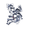
- Assembly
| Deposited unit | 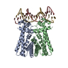
| ||||||||||||||||||
|---|---|---|---|---|---|---|---|---|---|---|---|---|---|---|---|---|---|---|---|
| 1 |
| ||||||||||||||||||
| Unit cell |
| ||||||||||||||||||
| Noncrystallographic symmetry (NCS) | NCS domain:
NCS domain segments: Component-ID: _ / Ens-ID: 1 / Beg auth comp-ID: SER / Beg label comp-ID: SER / End auth comp-ID: LEU / End label comp-ID: LEU / Refine code: _ / Auth seq-ID: 24 - 215 / Label seq-ID: 5 - 196
|
-Components
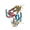
| #1: Protein | Mass: 21256.533 Da / Num. of mol.: 2 Source method: isolated from a genetically manipulated source Source: (gene. exp.) Mycobacterium tuberculosis (strain ATCC 25618 / H37Rv) (bacteria)Gene: Rv2506 / Production host: #2: DNA chain | | Mass: 6142.005 Da / Num. of mol.: 1 / Source method: obtained synthetically Source: (synth.) Mycobacterium tuberculosis H37Rv (bacteria)#3: DNA chain | | Mass: 6123.977 Da / Num. of mol.: 1 / Source method: obtained synthetically Source: (synth.) Mycobacterium tuberculosis H37Rv (bacteria) |
|---|
-Experimental details
-Experiment
| Experiment | Method: X-RAY DIFFRACTION / Number of used crystals: 1 |
|---|
- Sample preparation
| Crystal | Density Matthews: 3.16 Å3/Da / Density % sol: 61.1 % |
|---|---|
| Crystal grow | Temperature: 289 K / Method: vapor diffusion, sitting drop / pH: 5.5 Details: 0.2 M sodium acetate trihydrate, 0.1 M sodium citrate pH 5.5, 5% (w/v) PEG 4000. |
-Data collection
| Diffraction | Mean temperature: 100 K / Serial crystal experiment: N |
|---|---|
| Diffraction source | Source: SYNCHROTRON / Site: ESRF  / Beamline: ID23-1 / Wavelength: 0.97242 Å / Beamline: ID23-1 / Wavelength: 0.97242 Å |
| Detector | Type: ADSC QUANTUM 315 / Detector: CCD / Date: Mar 6, 2017 |
| Radiation | Protocol: SINGLE WAVELENGTH / Monochromatic (M) / Laue (L): M / Scattering type: x-ray |
| Radiation wavelength | Wavelength: 0.97242 Å / Relative weight: 1 |
| Reflection | Resolution: 3.15→62.03 Å / Num. obs: 12331 / % possible obs: 98.7 % / Redundancy: 4.3 % / Biso Wilson estimate: 74.4 Å2 / CC1/2: 0.999 / Rmerge(I) obs: 0.038 / Rpim(I) all: 0.02 / Net I/σ(I): 11.7 |
| Reflection shell | Resolution: 3.15→3.2 Å / Redundancy: 4.3 % / Rmerge(I) obs: 0.285 / Mean I/σ(I) obs: 1.8 / Num. unique obs: 597 / CC1/2: 0.982 / Rpim(I) all: 0.181 / % possible all: 99.5 |
- Processing
| Software |
| |||||||||||||||||||||||||||||||||||||||||||||||||||||||||||||||||||||||||||||||||||||||||||||||||||||||||||||||||||||||||||||
|---|---|---|---|---|---|---|---|---|---|---|---|---|---|---|---|---|---|---|---|---|---|---|---|---|---|---|---|---|---|---|---|---|---|---|---|---|---|---|---|---|---|---|---|---|---|---|---|---|---|---|---|---|---|---|---|---|---|---|---|---|---|---|---|---|---|---|---|---|---|---|---|---|---|---|---|---|---|---|---|---|---|---|---|---|---|---|---|---|---|---|---|---|---|---|---|---|---|---|---|---|---|---|---|---|---|---|---|---|---|---|---|---|---|---|---|---|---|---|---|---|---|---|---|---|---|---|
| Refinement | Method to determine structure: MOLECULAR REPLACEMENT Starting model: 6YJ2 Resolution: 3.15→62.03 Å / Cor.coef. Fo:Fc: 0.943 / Cor.coef. Fo:Fc free: 0.953 / SU B: 115.719 / SU ML: 0.701 / Cross valid method: THROUGHOUT / σ(F): 0 / ESU R Free: 0.52 / Stereochemistry target values: MAXIMUM LIKELIHOOD Details: HYDROGENS HAVE BEEN ADDED IN THE RIDING POSITIONS U VALUES : WITH TLS ADDED
| |||||||||||||||||||||||||||||||||||||||||||||||||||||||||||||||||||||||||||||||||||||||||||||||||||||||||||||||||||||||||||||
| Solvent computation | Ion probe radii: 0.8 Å / Shrinkage radii: 0.8 Å / VDW probe radii: 1.2 Å / Solvent model: MASK | |||||||||||||||||||||||||||||||||||||||||||||||||||||||||||||||||||||||||||||||||||||||||||||||||||||||||||||||||||||||||||||
| Displacement parameters | Biso max: 245.34 Å2 / Biso mean: 147.485 Å2 / Biso min: 105.52 Å2
| |||||||||||||||||||||||||||||||||||||||||||||||||||||||||||||||||||||||||||||||||||||||||||||||||||||||||||||||||||||||||||||
| Refinement step | Cycle: final / Resolution: 3.15→62.03 Å
| |||||||||||||||||||||||||||||||||||||||||||||||||||||||||||||||||||||||||||||||||||||||||||||||||||||||||||||||||||||||||||||
| Refine LS restraints |
| |||||||||||||||||||||||||||||||||||||||||||||||||||||||||||||||||||||||||||||||||||||||||||||||||||||||||||||||||||||||||||||
| Refine LS restraints NCS | Ens-ID: 1 / Number: 5844 / Refine-ID: X-RAY DIFFRACTION / Type: interatomic distance / Rms dev position: 0.09 Å / Weight position: 0.05
| |||||||||||||||||||||||||||||||||||||||||||||||||||||||||||||||||||||||||||||||||||||||||||||||||||||||||||||||||||||||||||||
| LS refinement shell | Resolution: 3.15→3.232 Å / Rfactor Rfree error: 0 / Total num. of bins used: 20
| |||||||||||||||||||||||||||||||||||||||||||||||||||||||||||||||||||||||||||||||||||||||||||||||||||||||||||||||||||||||||||||
| Refinement TLS params. | Method: refined / Refine-ID: X-RAY DIFFRACTION
| |||||||||||||||||||||||||||||||||||||||||||||||||||||||||||||||||||||||||||||||||||||||||||||||||||||||||||||||||||||||||||||
| Refinement TLS group |
|
