 Movie
Movie Controller

[English] 日本語
 Yorodumi
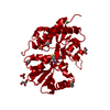
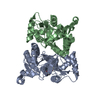
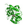
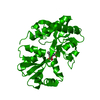
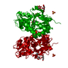
- PDB-6yk5: Structure of the AMPA receptor GluA2o ligand-binding domain (S1S2... -
+ Open data

- Basic information
| Entry | Database: PDB / ID: 6yk5 | ||||||
|---|---|---|---|---|---|---|---|
| Title | Structure of the AMPA receptor GluA2o ligand-binding domain (S1S2J) in complex with the compound (S)-1-(2'-Amino-2'-carboxyethyl)-5,7-dihydrofuro[3,4-d]- pyrimidine-2,4(1H,3H)-dione at resolution 1.15A | ||||||
 Components Components | Glutamate receptor 2 | ||||||
 Keywords Keywords | MEMBRANE PROTEIN / IONOTROPIC GLUTAMATE RECEPTOR / GLUA2O LIGAND-BINDING DOMAIN / BICYCLIC PYRIMIDINEDIONE ANALOGUE | ||||||
| Function / homology |  Function and homology information Function and homology informationspine synapse / dendritic spine neck / dendritic spine cytoplasm / dendritic spine head / cellular response to amine stimulus / Activation of AMPA receptors / ligand-gated monoatomic cation channel activity / perisynaptic space / Trafficking of GluR2-containing AMPA receptors / response to lithium ion ...spine synapse / dendritic spine neck / dendritic spine cytoplasm / dendritic spine head / cellular response to amine stimulus / Activation of AMPA receptors / ligand-gated monoatomic cation channel activity / perisynaptic space / Trafficking of GluR2-containing AMPA receptors / response to lithium ion / AMPA glutamate receptor activity / AMPA glutamate receptor clustering / kainate selective glutamate receptor activity / immunoglobulin binding / AMPA glutamate receptor complex / regulation of receptor recycling / extracellularly glutamate-gated ion channel activity / cellular response to glycine / ionotropic glutamate receptor complex / asymmetric synapse / Unblocking of NMDA receptors, glutamate binding and activation / glutamate receptor binding / conditioned place preference / positive regulation of synaptic transmission / response to fungicide / regulation of synaptic transmission, glutamatergic / extracellular ligand-gated monoatomic ion channel activity / cytoskeletal protein binding / glutamate-gated receptor activity / cellular response to brain-derived neurotrophic factor stimulus / regulation of long-term synaptic depression / somatodendritic compartment / glutamate-gated calcium ion channel activity / presynaptic active zone membrane / excitatory synapse / ionotropic glutamate receptor signaling pathway / ionotropic glutamate receptor binding / dendrite membrane / dendrite cytoplasm / ligand-gated monoatomic ion channel activity involved in regulation of presynaptic membrane potential / positive regulation of excitatory postsynaptic potential / dendritic shaft / SNARE binding / synaptic membrane / PDZ domain binding / establishment of protein localization / protein tetramerization / transmitter-gated monoatomic ion channel activity involved in regulation of postsynaptic membrane potential / synaptic transmission, glutamatergic / cerebral cortex development / receptor internalization / postsynaptic density membrane / modulation of chemical synaptic transmission / Schaffer collateral - CA1 synapse / terminal bouton / synaptic vesicle / long-term synaptic potentiation / amyloid-beta binding / synaptic vesicle membrane / growth cone / presynapse / signaling receptor activity / scaffold protein binding / presynaptic membrane / dendritic spine / chemical synaptic transmission / perikaryon / postsynaptic membrane / neuron projection / postsynaptic density / external side of plasma membrane / axon / neuronal cell body / synapse / dendrite / protein kinase binding / protein-containing complex binding / glutamatergic synapse / cell surface / endoplasmic reticulum / protein-containing complex / membrane / identical protein binding / plasma membrane Similarity search - Function | ||||||
| Biological species |  | ||||||
| Method |  X-RAY DIFFRACTION / SYNCHROTRON / MOLECULAR REPLACEMENT / Resolution: 1.15 Å X-RAY DIFFRACTION / SYNCHROTRON / MOLECULAR REPLACEMENT / Resolution: 1.15 Å | ||||||
 Authors Authors | Frydenvang, K. / Kastrup, J.S. | ||||||
 Citation Citation | Journal: Acs Chem Neurosci / Year: 2020 Title: Ionotropic Glutamate Receptor GluA2 in Complex with Bicyclic Pyrimidinedione-Based Compounds: When Small Compound Modifications Have Distinct Effects on Binding Interactions. Authors: Frydenvang, K. / Pickering, D.S. / Kshirsagar, G.U. / Chemi, G. / Gemma, S. / Sprogoe, D. / Kærn, A.M. / Brogi, S. / Campiani, G. / Butini, S. / Kastrup, J.S. | ||||||
| History |
|
- Structure visualization
| Structure viewer | Molecule: MolmilJmol/JSmol |
|---|
- Downloads & links
-Download
| PDBx/mmCIF format | 6yk5.cif.gz | 146.3 KB | Display | PDBx/mmCIF format |
|---|---|---|---|---|
| PDB format | pdb6yk5.ent.gz | 112.9 KB | Display | PDB format |
| PDBx/mmJSON format | 6yk5.json.gz | Tree view | PDBx/mmJSON format | |
| Others |  Other downloads Other downloads |
-Validation report
| Arichive directory | https://data.pdbj.org/pub/pdb/validation_reports/yk/6yk5ftp://data.pdbj.org/pub/pdb/validation_reports/yk/6yk5 | HTTPS FTP |
|---|
-Related structure data
| Related structure data |  6yk2C  6yk3C 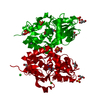 6yk4C  6yk6C 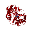 1syhS S: Starting model for refinement C: citing same article ( |
|---|---|
| Similar structure data |


-Links
 PDBj
PDBj


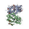
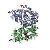
- Assembly
| Deposited unit | 
| ||||||||
|---|---|---|---|---|---|---|---|---|---|
| 1 | 
| ||||||||
| Unit cell |
| ||||||||
| Components on special symmetry positions |
|
-Components
-Protein , 1 types, 1 molecules A
| #1: Protein | Mass: 29278.732 Da / Num. of mol.: 1 Source method: isolated from a genetically manipulated source Details: THE PROTEIN CRYSTALLIZED IS THE EXTRACELLULAR LIGAND-BINDING DOMAIN OF GLUA2O. TRANSMEMBRANE REGIONS WERE GENETICALLY REMOVED AND REPLACED WITH A GLY-THR LINKER (RESIDUE 118-119). THE ...Details: THE PROTEIN CRYSTALLIZED IS THE EXTRACELLULAR LIGAND-BINDING DOMAIN OF GLUA2O. TRANSMEMBRANE REGIONS WERE GENETICALLY REMOVED AND REPLACED WITH A GLY-THR LINKER (RESIDUE 118-119). THE SEQUENCE MATCHES DISCONTINOUSLY WITH THE REFERENCE DATABASE (UNP RESIDUES 413-527 AND 653-797, NUMBERING WITH SIGNAL PEPTIDE OF 21 AMINO ACIDS). THE TWO FIRST RESIDUES (GLY, ALA) ARE CLONING REMNANTS. Source: (gene. exp.)  |
|---|
-Non-polymers , 6 types, 459 molecules 


| #2: Chemical | ChemComp-GOL /  Mass: 92.094 Da / Num. of mol.: 4 / Source method: obtained synthetically / Formula: C3H8O3 Mass: 92.094 Da / Num. of mol.: 4 / Source method: obtained synthetically / Formula: C3H8O3#3: Chemical | ChemComp-SO4 /  Mass: 96.063 Da / Num. of mol.: 6 / Source method: obtained synthetically / Formula: SO4 Mass: 96.063 Da / Num. of mol.: 6 / Source method: obtained synthetically / Formula: SO4#4: Chemical | ChemComp-LI / |  Mass: 6.941 Da / Num. of mol.: 1 / Source method: obtained synthetically / Formula: Li Mass: 6.941 Da / Num. of mol.: 1 / Source method: obtained synthetically / Formula: Li#5: Chemical | ChemComp-PVK / ( |  Mass: 241.201 Da / Num. of mol.: 1 / Source method: obtained synthetically / Formula: C9H11N3O5 / Feature type: SUBJECT OF INVESTIGATION Mass: 241.201 Da / Num. of mol.: 1 / Source method: obtained synthetically / Formula: C9H11N3O5 / Feature type: SUBJECT OF INVESTIGATION#6: Chemical | ChemComp-CL / |  Mass: 35.453 Da / Num. of mol.: 1 / Source method: obtained synthetically / Formula: Cl Mass: 35.453 Da / Num. of mol.: 1 / Source method: obtained synthetically / Formula: Cl#7: Water | ChemComp-HOH / | Mass: 18.015 Da / Num. of mol.: 446 / Source method: isolated from a natural source / Formula: H2O |
|---|
-Details
| Has ligand of interest | Y |
|---|---|
| Has protein modification | Y |
-Experimental details
-Experiment
| Experiment | Method: X-RAY DIFFRACTION / Number of used crystals: 1 |
|---|
- Sample preparation
| Crystal | Density Matthews: 2.36 Å3/Da / Density % sol: 47.83 % |
|---|---|
| Crystal grow | Temperature: 279 K / Method: vapor diffusion, hanging drop / pH: 5.5 / Details: PEG4000, lithium sulfate, acetate |
-Data collection
| Diffraction | Mean temperature: 100 K / Serial crystal experiment: N | |||||||||||||||||||||||||||||||||||||||||||||||||||||||||||||||||||||||||||||||||||||||||||||||||||||||||||||||||||||||||
|---|---|---|---|---|---|---|---|---|---|---|---|---|---|---|---|---|---|---|---|---|---|---|---|---|---|---|---|---|---|---|---|---|---|---|---|---|---|---|---|---|---|---|---|---|---|---|---|---|---|---|---|---|---|---|---|---|---|---|---|---|---|---|---|---|---|---|---|---|---|---|---|---|---|---|---|---|---|---|---|---|---|---|---|---|---|---|---|---|---|---|---|---|---|---|---|---|---|---|---|---|---|---|---|---|---|---|---|---|---|---|---|---|---|---|---|---|---|---|---|---|---|---|
| Diffraction source | Source: SYNCHROTRON / Site: MAX II  / Beamline: I911-3 / Wavelength: 1 Å / Beamline: I911-3 / Wavelength: 1 Å | |||||||||||||||||||||||||||||||||||||||||||||||||||||||||||||||||||||||||||||||||||||||||||||||||||||||||||||||||||||||||
| Detector | Type: MARMOSAIC 225 mm CCD / Detector: CCD / Date: May 14, 2013 | |||||||||||||||||||||||||||||||||||||||||||||||||||||||||||||||||||||||||||||||||||||||||||||||||||||||||||||||||||||||||
| Radiation | Protocol: SINGLE WAVELENGTH / Monochromatic (M) / Laue (L): M / Scattering type: x-ray | |||||||||||||||||||||||||||||||||||||||||||||||||||||||||||||||||||||||||||||||||||||||||||||||||||||||||||||||||||||||||
| Radiation wavelength | Wavelength: 1 Å / Relative weight: 1 | |||||||||||||||||||||||||||||||||||||||||||||||||||||||||||||||||||||||||||||||||||||||||||||||||||||||||||||||||||||||||
| Reflection | Resolution: 1.15→28.212 Å / Num. all: 97655 / Num. obs: 97655 / % possible obs: 98.6 % / Redundancy: 4.6 % / Biso Wilson estimate: 7.97 Å2 / Rpim(I) all: 0.032 / Rrim(I) all: 0.069 / Rsym value: 0.061 / Net I/av σ(I): 7.6 / Net I/σ(I): 14.2 / Num. measured all: 450981 | |||||||||||||||||||||||||||||||||||||||||||||||||||||||||||||||||||||||||||||||||||||||||||||||||||||||||||||||||||||||||
| Reflection shell | Diffraction-ID: 1
|
- Processing
| Software |
| ||||||||||||||||||||||||||||||||||||||||||||||||||||||||||||||||||||||||||||||||||||||||||||||||||||||||||||||||||||||||||||||||||||||||||||||||||||||||||||||||||||||||||||||||||||||||||
|---|---|---|---|---|---|---|---|---|---|---|---|---|---|---|---|---|---|---|---|---|---|---|---|---|---|---|---|---|---|---|---|---|---|---|---|---|---|---|---|---|---|---|---|---|---|---|---|---|---|---|---|---|---|---|---|---|---|---|---|---|---|---|---|---|---|---|---|---|---|---|---|---|---|---|---|---|---|---|---|---|---|---|---|---|---|---|---|---|---|---|---|---|---|---|---|---|---|---|---|---|---|---|---|---|---|---|---|---|---|---|---|---|---|---|---|---|---|---|---|---|---|---|---|---|---|---|---|---|---|---|---|---|---|---|---|---|---|---|---|---|---|---|---|---|---|---|---|---|---|---|---|---|---|---|---|---|---|---|---|---|---|---|---|---|---|---|---|---|---|---|---|---|---|---|---|---|---|---|---|---|---|---|---|---|---|---|---|
| Refinement | Method to determine structure: MOLECULAR REPLACEMENT Starting model: 1SYH Resolution: 1.15→26.778 Å / SU ML: 0.08 / Cross valid method: THROUGHOUT / σ(F): 1.35 / Phase error: 9.7
| ||||||||||||||||||||||||||||||||||||||||||||||||||||||||||||||||||||||||||||||||||||||||||||||||||||||||||||||||||||||||||||||||||||||||||||||||||||||||||||||||||||||||||||||||||||||||||
| Solvent computation | Shrinkage radii: 0.9 Å / VDW probe radii: 1.11 Å | ||||||||||||||||||||||||||||||||||||||||||||||||||||||||||||||||||||||||||||||||||||||||||||||||||||||||||||||||||||||||||||||||||||||||||||||||||||||||||||||||||||||||||||||||||||||||||
| Displacement parameters | Biso max: 55.22 Å2 / Biso mean: 12.6496 Å2 / Biso min: 4.09 Å2 | ||||||||||||||||||||||||||||||||||||||||||||||||||||||||||||||||||||||||||||||||||||||||||||||||||||||||||||||||||||||||||||||||||||||||||||||||||||||||||||||||||||||||||||||||||||||||||
| Refinement step | Cycle: final / Resolution: 1.15→26.778 Å
| ||||||||||||||||||||||||||||||||||||||||||||||||||||||||||||||||||||||||||||||||||||||||||||||||||||||||||||||||||||||||||||||||||||||||||||||||||||||||||||||||||||||||||||||||||||||||||
| LS refinement shell | Refine-ID: X-RAY DIFFRACTION / Rfactor Rfree error: 0
|
