 Movie
Movie Controller

[English] 日本語
 Yorodumi
- PDB-6yjt: Crystal structure of MGAT5 (alpha-1,6-mannosylglycoprotein 6-beta... -
+ Open data

- Basic information
| Entry | Database: PDB / ID: 6yjt | ||||||
|---|---|---|---|---|---|---|---|
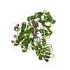
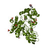
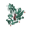
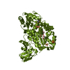
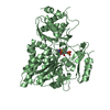
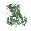
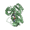
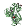
| Title | Crystal structure of MGAT5 (alpha-1,6-mannosylglycoprotein 6-beta-N-acetylglucosaminyltransferase V) luminal domain with a Lys329-Ile345 loop truncation, in complex with UDP | ||||||
 Components Components | Alpha-1,6-mannosylglycoprotein 6-beta-N-acetylglucosaminyltransferase A | ||||||
 Keywords Keywords | TRANSFERASE / Carbohydrate / Enzyme / N-glycosylation / GlcNAc | ||||||
| Function / homology |  Function and homology information Function and homology informationalpha-1,6-mannosyl-glycoprotein 6-beta-N-acetylglucosaminyltransferase / alpha-1,6-mannosylglycoprotein 6-beta-N-acetylglucosaminyltransferase activity / N-Glycan antennae elongation / positive regulation of receptor signaling pathway via STAT / : / protein N-linked glycosylation / protein phosphatase inhibitor activity / manganese ion binding / Maturation of spike protein / viral protein processing ...alpha-1,6-mannosyl-glycoprotein 6-beta-N-acetylglucosaminyltransferase / alpha-1,6-mannosylglycoprotein 6-beta-N-acetylglucosaminyltransferase activity / N-Glycan antennae elongation / positive regulation of receptor signaling pathway via STAT / : / protein N-linked glycosylation / protein phosphatase inhibitor activity / manganese ion binding / Maturation of spike protein / viral protein processing / positive regulation of cell migration / Golgi membrane / Golgi apparatus / extracellular exosome / membrane Similarity search - Function | ||||||
| Biological species |  Homo sapiens (human) Homo sapiens (human) | ||||||
| Method |  X-RAY DIFFRACTION / SYNCHROTRON / MOLECULAR REPLACEMENT / Resolution: 1.7 Å X-RAY DIFFRACTION / SYNCHROTRON / MOLECULAR REPLACEMENT / Resolution: 1.7 Å | ||||||
 Authors Authors | Wu, L. / Darby, J.F. / Gilio, A.K. / Davies, G.J. | ||||||
| Funding support |  United Kingdom, 1items United Kingdom, 1items
| ||||||
 Citation Citation | Journal: Acs Catalysis / Year: 2020 Title: Substrate Engagement and Catalytic Mechanisms of N-Acetylglucosaminyltransferase V Authors: Darby, J.F. / Gilio, A.K. / Piniello, B. / Roth, C. / Blagova, E. / Rovira, C. / Hubbard, R.E. / Davies, G.J. / Wu, L. | ||||||
| History |
|
- Structure visualization
| Structure viewer | Molecule: MolmilJmol/JSmol |
|---|
- Downloads & links
-Download
| PDBx/mmCIF format | 6yjt.cif.gz | 425.8 KB | Display | PDBx/mmCIF format |
|---|---|---|---|---|
| PDB format | pdb6yjt.ent.gz | Display | PDB format | |
| PDBx/mmJSON format | 6yjt.json.gz | Tree view | PDBx/mmJSON format | |
| Others |  Other downloads Other downloads |
-Validation report
| Arichive directory | https://data.pdbj.org/pub/pdb/validation_reports/yj/6yjtftp://data.pdbj.org/pub/pdb/validation_reports/yj/6yjt | HTTPS FTP |
|---|
-Related structure data
| Related structure data | 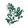 6yjqC 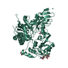 6yjrC 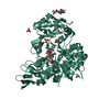 6yjsC  6yjuC  6yjvC 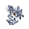 5zicS S: Starting model for refinement C: citing same article ( |
|---|---|
| Similar structure data |

-Links
 PDBj
PDBj


- Assembly
| Deposited unit | 
| ||||||||
|---|---|---|---|---|---|---|---|---|---|
| 1 | 
| ||||||||
| 2 | 
| ||||||||
| Unit cell |
| ||||||||
| Noncrystallographic symmetry (NCS) | NCS domain: (Details: Chains AAA BBB) |
-Components
-Protein / Sugars , 2 types, 4 molecules AAABBB
| #1: Protein | Mass: 58996.906 Da / Num. of mol.: 2 Source method: isolated from a genetically manipulated source Source: (gene. exp.) Homo sapiens (human) / Gene: MGAT5, GGNT5 / Production host:  Trichoplusia ni (cabbage looper) Trichoplusia ni (cabbage looper)References: UniProt: Q09328, alpha-1,6-mannosyl-glycoprotein 6-beta-N-acetylglucosaminyltransferase #4: Sugar |  Type: D-saccharide, beta linking / Mass: 221.208 Da / Num. of mol.: 2 Type: D-saccharide, beta linking / Mass: 221.208 Da / Num. of mol.: 2Source method: isolated from a genetically manipulated source Formula: C8H15NO6 |
|---|
-Non-polymers , 4 types, 346 molecules 


| #2: Chemical |  Type: RNA linking / Mass: 404.161 Da / Num. of mol.: 2 / Source method: obtained synthetically / Formula: C9H14N2O12P2 / Feature type: SUBJECT OF INVESTIGATION / Comment: UDP*YM Type: RNA linking / Mass: 404.161 Da / Num. of mol.: 2 / Source method: obtained synthetically / Formula: C9H14N2O12P2 / Feature type: SUBJECT OF INVESTIGATION / Comment: UDP*YM#3: Chemical | ChemComp-EDO /  Mass: 62.068 Da / Num. of mol.: 8 / Source method: obtained synthetically / Formula: C2H6O2 Mass: 62.068 Da / Num. of mol.: 8 / Source method: obtained synthetically / Formula: C2H6O2#5: Chemical | ChemComp-SO4 /  Mass: 96.063 Da / Num. of mol.: 6 / Source method: obtained synthetically / Formula: SO4 Mass: 96.063 Da / Num. of mol.: 6 / Source method: obtained synthetically / Formula: SO4#6: Water | ChemComp-HOH / | Mass: 18.015 Da / Num. of mol.: 330 / Source method: isolated from a natural source / Formula: H2O |
|---|
-Details
| Has ligand of interest | Y |
|---|---|
| Has protein modification | Y |
-Experimental details
-Experiment
| Experiment | Method: X-RAY DIFFRACTION / Number of used crystals: 1 |
|---|
- Sample preparation
| Crystal | Density Matthews: 2.25 Å3/Da / Density % sol: 45.43 % |
|---|---|
| Crystal grow | Temperature: 293 K / Method: vapor diffusion, sitting drop Details: 0.1 M HEPES pH 8.0, 0.3 M Li2SO4, 30 % (w/v) PEG 3350, 10 % (v/v) ethylene glycol |
-Data collection
| Diffraction | Mean temperature: 100 K / Serial crystal experiment: N |
|---|---|
| Diffraction source | Source: SYNCHROTRON / Site: Diamond / Beamline: I03 / Wavelength: 0.97625 Å |
| Detector | Type: DECTRIS PILATUS 6M / Detector: PIXEL / Date: Apr 8, 2019 |
| Radiation | Protocol: SINGLE WAVELENGTH / Monochromatic (M) / Laue (L): M / Scattering type: x-ray |
| Radiation wavelength | Wavelength: 0.97625 Å / Relative weight: 1 |
| Reflection | Resolution: 1.7→43.43 Å / Num. obs: 108811 / % possible obs: 96 % / Redundancy: 3.6 % / CC1/2: 0.999 / Rmerge(I) obs: 0.047 / Rpim(I) all: 0.03 / Net I/σ(I): 9.9 |
| Reflection shell | Resolution: 1.7→1.74 Å / Rmerge(I) obs: 1.346 / Mean I/σ(I) obs: 1 / Num. unique obs: 8076 / CC1/2: 0.562 / Rpim(I) all: 0.804 |
- Processing
| Software |
| |||||||||||||||||||||||||||||||||||||||||||||||||||||||||||||||||||||||||||||||||||||||||||||||||||||||||||||||||||||||||||||||||||||||||||||||||||||||||||
|---|---|---|---|---|---|---|---|---|---|---|---|---|---|---|---|---|---|---|---|---|---|---|---|---|---|---|---|---|---|---|---|---|---|---|---|---|---|---|---|---|---|---|---|---|---|---|---|---|---|---|---|---|---|---|---|---|---|---|---|---|---|---|---|---|---|---|---|---|---|---|---|---|---|---|---|---|---|---|---|---|---|---|---|---|---|---|---|---|---|---|---|---|---|---|---|---|---|---|---|---|---|---|---|---|---|---|---|---|---|---|---|---|---|---|---|---|---|---|---|---|---|---|---|---|---|---|---|---|---|---|---|---|---|---|---|---|---|---|---|---|---|---|---|---|---|---|---|---|---|---|---|---|---|---|---|---|
| Refinement | Method to determine structure: MOLECULAR REPLACEMENT Starting model: 5zic Resolution: 1.7→43.43 Å / Cor.coef. Fo:Fc: 0.976 / Cor.coef. Fo:Fc free: 0.953 / SU B: 3.645 / SU ML: 0.116 / Cross valid method: FREE R-VALUE / ESU R: 0.114 / ESU R Free: 0.112 Details: Hydrogens have been added in their riding positions
| |||||||||||||||||||||||||||||||||||||||||||||||||||||||||||||||||||||||||||||||||||||||||||||||||||||||||||||||||||||||||||||||||||||||||||||||||||||||||||
| Solvent computation | Ion probe radii: 0.8 Å / Shrinkage radii: 0.8 Å / VDW probe radii: 1.2 Å | |||||||||||||||||||||||||||||||||||||||||||||||||||||||||||||||||||||||||||||||||||||||||||||||||||||||||||||||||||||||||||||||||||||||||||||||||||||||||||
| Displacement parameters | Biso mean: 54.886 Å2
| |||||||||||||||||||||||||||||||||||||||||||||||||||||||||||||||||||||||||||||||||||||||||||||||||||||||||||||||||||||||||||||||||||||||||||||||||||||||||||
| Refinement step | Cycle: LAST / Resolution: 1.7→43.43 Å
| |||||||||||||||||||||||||||||||||||||||||||||||||||||||||||||||||||||||||||||||||||||||||||||||||||||||||||||||||||||||||||||||||||||||||||||||||||||||||||
| Refine LS restraints |
| |||||||||||||||||||||||||||||||||||||||||||||||||||||||||||||||||||||||||||||||||||||||||||||||||||||||||||||||||||||||||||||||||||||||||||||||||||||||||||
| LS refinement shell |
|
