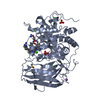
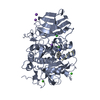
Entry Database : PDB / ID : 6yd7Title X-ray structure of furin in complex with the canavanine-based inhibitor 4-guanidinomethyl-phenylacetyl-Arg-Tle-Canavanine-Amba 4-guanidinomethyl-phenylacetyl-Arg-Tle-Canavanine-Amba Furin Keywords / / / / Function / homology Function Domain/homology Component
/ / / / / / / / / / / / / / / / / / / / / / / / / / / / / / / / / / / / / / / / / / / / / / / / / / / / / / / / / / / / / / / / / / / / / / / / / / / / / / / / / / / / / / / / / / / / / / / / / / / / / / / / / / / / / / / / / / / / / / / / / / Biological species Homo sapiens (human)synthetic construct (others) Method / / / Resolution : 1.8 Å Authors Dahms, S.O. Funding support Organization Grant number Country Austrian Science Fund M 2730
Journal : Acs Med.Chem.Lett. / Year : 2021Title : The Basicity Makes the Difference: Improved Canavanine-Derived Inhibitors of the Proprotein Convertase Furin.Authors : Lam van, T.V. / Heindl, M.R. / Schlutt, C. / Bottcher-Friebertshauser, E. / Bartenschlager, R. / Klebe, G. / Brandstetter, H. / Dahms, S.O. / Steinmetzer, T. History Deposition Mar 20, 2020 Deposition site / Processing site Revision 1.0 Feb 17, 2021 Provider / Type Revision 1.1 Mar 31, 2021 Group / Category / citation_authorItem _citation.journal_volume / _citation.page_first ... _citation.journal_volume / _citation.page_first / _citation.page_last / _citation.pdbx_database_id_PubMed / _citation.title / _citation_author.name Revision 2.0 Oct 19, 2022 Group Advisory / Atomic model ... Advisory / Atomic model / Data collection / Database references / Derived calculations / Non-polymer description / Polymer sequence / Source and taxonomy / Structure summary Category atom_site / chem_comp ... atom_site / chem_comp / database_2 / entity / entity_poly / entity_poly_seq / entity_src_gen / pdbx_entity_nonpoly / pdbx_entity_src_syn / pdbx_nonpoly_scheme / pdbx_poly_seq_scheme / pdbx_struct_assembly / pdbx_struct_assembly_gen / pdbx_struct_assembly_prop / pdbx_struct_conn_angle / pdbx_struct_sheet_hbond / pdbx_struct_special_symmetry / pdbx_unobs_or_zero_occ_atoms / struct_asym / struct_conn / struct_ref / struct_ref_seq / struct_sheet / struct_sheet_order / struct_sheet_range / struct_site / struct_site_gen Item _atom_site.B_iso_or_equiv / _atom_site.Cartn_x ... _atom_site.B_iso_or_equiv / _atom_site.Cartn_x / _atom_site.Cartn_y / _atom_site.Cartn_z / _atom_site.auth_asym_id / _atom_site.auth_atom_id / _atom_site.auth_comp_id / _atom_site.auth_seq_id / _atom_site.group_PDB / _atom_site.label_alt_id / _atom_site.label_asym_id / _atom_site.label_atom_id / _atom_site.label_comp_id / _atom_site.label_entity_id / _atom_site.label_seq_id / _atom_site.occupancy / _atom_site.type_symbol / _chem_comp.formula / _chem_comp.formula_weight / _chem_comp.id / _chem_comp.mon_nstd_flag / _chem_comp.name / _chem_comp.pdbx_synonyms / _chem_comp.type / _database_2.pdbx_DOI / _database_2.pdbx_database_accession / _entity_src_gen.gene_src_common_name / _pdbx_struct_assembly.details / _pdbx_struct_assembly.method_details / _pdbx_struct_assembly.oligomeric_count / _pdbx_struct_assembly.oligomeric_details / _pdbx_struct_assembly_gen.asym_id_list / _pdbx_struct_assembly_prop.value / _pdbx_struct_conn_angle.ptnr1_auth_seq_id / _pdbx_struct_conn_angle.ptnr1_label_asym_id / _pdbx_struct_conn_angle.ptnr2_label_asym_id / _pdbx_struct_conn_angle.ptnr3_auth_seq_id / _pdbx_struct_conn_angle.ptnr3_label_asym_id / _pdbx_struct_special_symmetry.auth_seq_id / _pdbx_struct_special_symmetry.label_asym_id / _struct_conn.pdbx_leaving_atom_flag / _struct_conn.ptnr1_auth_asym_id / _struct_conn.ptnr1_auth_comp_id / _struct_conn.ptnr1_label_asym_id / _struct_conn.ptnr1_label_atom_id / _struct_conn.ptnr1_label_comp_id / _struct_conn.ptnr1_label_seq_id / _struct_conn.ptnr2_auth_asym_id / _struct_conn.ptnr2_auth_comp_id / _struct_conn.ptnr2_auth_seq_id / _struct_conn.ptnr2_label_asym_id / _struct_conn.ptnr2_label_comp_id / _struct_conn.ptnr2_label_seq_id / _struct_site.pdbx_auth_asym_id / _struct_site.pdbx_auth_comp_id / _struct_site_gen.auth_asym_id / _struct_site_gen.auth_comp_id / _struct_site_gen.auth_seq_id / _struct_site_gen.label_asym_id / _struct_site_gen.label_comp_id / _struct_site_gen.label_seq_id Revision 3.0 Nov 15, 2023 Group / Data collection / Category / chem_comp_atom / chem_comp_bond / Item / _atom_site.label_atom_idRevision 3.1 Jan 31, 2024 Group / Category
Show all Show less
 Movie
Movie Controller

 Yorodumi
 Open data
 Basic information
 Components
 Keywords
 Function and homology information
 Homo sapiens (human)
 X-RAY DIFFRACTION /
  Authors
 Austria, 1items
  Citation
 Structure visualization
 Downloads & links
 Other downloads
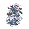


 PDBj
PDBj


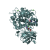
 Assembly
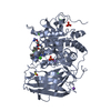


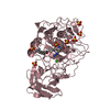
 Mass: 40.078 Da / Num. of mol.: 3 / Source method: obtained synthetically / Formula: Ca
 Mass: 22.990 Da / Num. of mol.: 3 / Source method: obtained synthetically / Formula: Na
 Mass: 35.453 Da / Num. of mol.: 1 / Source method: obtained synthetically / Formula: Cl
 Mass: 94.971 Da / Num. of mol.: 2 / Source method: obtained synthetically / Formula: PO4
 Mass: 78.133 Da / Num. of mol.: 1 / Source method: obtained synthetically / Formula: C2H6OS / Comment: DMSO, precipitant*YM
 Sample preparation
 / Beamline: 14.2 / Wavelength: 0.9184 Å
 Processing