 Movie
Movie Controller

[English] 日本語
 Yorodumi
- PDB-6y93: Crystal structure of the DNA-binding domain of the Nucleoid Occlu... -
+ Open data

- Basic information
| Entry | Database: PDB / ID: 6y93 | ||||||||||||||||||
|---|---|---|---|---|---|---|---|---|---|---|---|---|---|---|---|---|---|---|---|
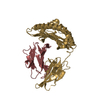
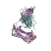
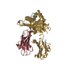
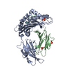
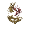
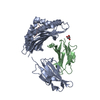
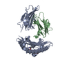
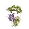
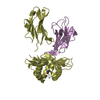
| Title | Crystal structure of the DNA-binding domain of the Nucleoid Occlusion Factor (Noc) complexed to the Noc-binding site (NBS) | ||||||||||||||||||
 Components Components |
| ||||||||||||||||||
 Keywords Keywords | DNA BINDING PROTEIN / chromosome segregation / chromosome maintenance / protein-DNA recognition / evolution / DNA-binding protein | ||||||||||||||||||
| Function / homology |  Function and homology information Function and homology informationpositive regulation of sporulation resulting in formation of a cellular spore / division septum assembly / nucleoid / chromosome segregation / chromosome / DNA binding / cytoplasm Similarity search - Function | ||||||||||||||||||
| Biological species |  synthetic construct (others) | ||||||||||||||||||
| Method |  X-RAY DIFFRACTION / SYNCHROTRON / MOLECULAR REPLACEMENT / Resolution: 2.23 Å X-RAY DIFFRACTION / SYNCHROTRON / MOLECULAR REPLACEMENT / Resolution: 2.23 Å | ||||||||||||||||||
 Authors Authors | Jalal, A.S.B. / Tran, N.T. / Stevenson, C.E.M. / Chan, E. / Lo, R. / Tan, X. / Noy, A. / Lawson, D.M. / Le, T.B.K. | ||||||||||||||||||
| Funding support |  United Kingdom, 5items United Kingdom, 5items
| ||||||||||||||||||
 Citation Citation | Journal: Cell Rep / Year: 2020 Title: Diversification of DNA-Binding Specificity by Permissive and Specificity-Switching Mutations in the ParB/Noc Protein Family. Authors: Jalal, A.S.B. / Tran, N.T. / Stevenson, C.E. / Chan, E.W. / Lo, R. / Tan, X. / Noy, A. / Lawson, D.M. / Le, T.B.K. | ||||||||||||||||||
| History |
|
- Structure visualization
| Structure viewer | Molecule: MolmilJmol/JSmol |
|---|
- Downloads & links
-Download
| PDBx/mmCIF format | 6y93.cif.gz | 148 KB | Display | PDBx/mmCIF format |
|---|---|---|---|---|
| PDB format | pdb6y93.ent.gz | 114.2 KB | Display | PDB format |
| PDBx/mmJSON format | 6y93.json.gz | Tree view | PDBx/mmJSON format | |
| Others |  Other downloads Other downloads |
-Validation report
| Summary document | 6y93_validation.pdf.gz | 455 KB | Display | wwPDB validaton report |
|---|---|---|---|---|
| Full document | 6y93_full_validation.pdf.gz | 466.5 KB | Display | |
| Data in XML | 6y93_validation.xml.gz | 10.4 KB | Display | |
| Data in CIF | 6y93_validation.cif.gz | 13.4 KB | Display | |
| Arichive directory | https://data.pdbj.org/pub/pdb/validation_reports/y9/6y93ftp://data.pdbj.org/pub/pdb/validation_reports/y9/6y93 | HTTPS FTP |
-Related structure data
| Related structure data |  6s6hSC S: Starting model for refinement C: citing same article ( |
|---|---|
| Similar structure data |

-Links
 PDBj
PDBj


- Assembly
| Deposited unit | 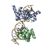
| ||||||||||||||||||||||||||||||||||||||||||||||||||||||||||||||||||||
|---|---|---|---|---|---|---|---|---|---|---|---|---|---|---|---|---|---|---|---|---|---|---|---|---|---|---|---|---|---|---|---|---|---|---|---|---|---|---|---|---|---|---|---|---|---|---|---|---|---|---|---|---|---|---|---|---|---|---|---|---|---|---|---|---|---|---|---|---|---|
| 1 |
| ||||||||||||||||||||||||||||||||||||||||||||||||||||||||||||||||||||
| Unit cell |
| ||||||||||||||||||||||||||||||||||||||||||||||||||||||||||||||||||||
| Noncrystallographic symmetry (NCS) | NCS domain:
NCS domain segments: Component-ID: _ / Refine code: _
NCS ensembles :
|
-Components
| #1: Protein | Mass: 16806.496 Da / Num. of mol.: 2 Source method: isolated from a genetically manipulated source Details: The expressed protein comprised residues 111-242 of the wild-type sequence with an additional N-terminal Met residue and a C-terminal nickel affinity tag of sequence KLAAALEHHHHHH from the ...Details: The expressed protein comprised residues 111-242 of the wild-type sequence with an additional N-terminal Met residue and a C-terminal nickel affinity tag of sequence KLAAALEHHHHHH from the pET21b expression plasmid Source: (gene. exp.) Gene: noc, yyaA, BSU40990 / Production host: #2: DNA chain | Mass: 6751.378 Da / Num. of mol.: 2 / Source method: obtained synthetically Details: Chains C and D form a symmetrical DNA duplex which is similar to the Noc Binding Site (NBS) sequence from Bacillus subtilis without 5' phosphates Source: (synth.) synthetic construct (others) |
|---|
-Experimental details
-Experiment
| Experiment | Method: X-RAY DIFFRACTION / Number of used crystals: 1 |
|---|
- Sample preparation
| Crystal | Density Matthews: 3.94 Å3/Da / Density % sol: 68.8 % |
|---|---|
| Crystal grow | Temperature: 293 K / Method: vapor diffusion, sitting drop / pH: 8 / Details: NULL |
-Data collection
| Diffraction | Mean temperature: 100 K / Serial crystal experiment: N | ||||||||||||||||||||||||
|---|---|---|---|---|---|---|---|---|---|---|---|---|---|---|---|---|---|---|---|---|---|---|---|---|---|
| Diffraction source | Source: SYNCHROTRON / Site: Diamond / Beamline: I03 / Wavelength: 0.9762 Å | ||||||||||||||||||||||||
| Detector | Type: DECTRIS EIGER2 XE 16M / Detector: PIXEL / Date: Apr 27, 2019 | ||||||||||||||||||||||||
| Radiation | Protocol: SINGLE WAVELENGTH / Monochromatic (M) / Laue (L): M / Scattering type: x-ray | ||||||||||||||||||||||||
| Radiation wavelength | Wavelength: 0.9762 Å / Relative weight: 1 | ||||||||||||||||||||||||
| Reflection | Resolution: 2.23→72.3 Å / Num. obs: 10830 / % possible obs: 38.1 % / Redundancy: 13.1 % / CC1/2: 1 / Rmerge(I) obs: 0.108 / Rpim(I) all: 0.031 / Rrim(I) all: 0.112 / Net I/σ(I): 9.3 | ||||||||||||||||||||||||
| Reflection shell | Diffraction-ID: 1
|
- Processing
| Software |
| |||||||||||||||||||||||||||||||||||||||||||||||||||||||||||||||||||||||||||||||||||||||||||||||||||||||||||||||||||||||||||||
|---|---|---|---|---|---|---|---|---|---|---|---|---|---|---|---|---|---|---|---|---|---|---|---|---|---|---|---|---|---|---|---|---|---|---|---|---|---|---|---|---|---|---|---|---|---|---|---|---|---|---|---|---|---|---|---|---|---|---|---|---|---|---|---|---|---|---|---|---|---|---|---|---|---|---|---|---|---|---|---|---|---|---|---|---|---|---|---|---|---|---|---|---|---|---|---|---|---|---|---|---|---|---|---|---|---|---|---|---|---|---|---|---|---|---|---|---|---|---|---|---|---|---|---|---|---|---|
| Refinement | Method to determine structure: MOLECULAR REPLACEMENT Starting model: 6S6H Resolution: 2.23→72.3 Å / Cor.coef. Fo:Fc: 0.949 / Cor.coef. Fo:Fc free: 0.921 / SU ML: 0.385 / SU R Cruickshank DPI: 0.3856 / Cross valid method: THROUGHOUT / σ(F): 0 / ESU R Free: 0.441 Details: HYDROGENS HAVE BEEN ADDED IN THE RIDING POSITIONS U VALUES: WITH TLS ADDED This structure was refined after applying an anisotropic correction to the data with STARANISO. In line with the ...Details: HYDROGENS HAVE BEEN ADDED IN THE RIDING POSITIONS U VALUES: WITH TLS ADDED This structure was refined after applying an anisotropic correction to the data with STARANISO. In line with the recommendations of the PDB and the STARANISO webpages, the uploaded data cif file contains three data blocks: 1. the STARANISO corrected data that was used to refine the structure, 2. the full uncorrected dataset with no resolution cut-off applied, 3. the output from refmac5 with filled-in data removed to leave only the reflections that were observed within the ellipsoidal resolution cut-off. The latter contains blurred map coefficients that should enable reproduction of the maps that were used to evaluate the model. N.B. The anisotropic correction leads to poor completeness in the higher spherical resolution shells resulting in maps that appear to be at lower resolution than the 2.23 Ang maximum resolution of the corrected data. Since the Rfree and RSRZ scores which evaluate the fit of the model to the data are dependent on the maximum reported resolution, they are inevitably poor for this model. Also note that the map coefficients automatically generated by the deposition process have unfortunately re-instated the above mentioned filled-in reflections. Thus, these will produce maps with an element of model bias i.e. they will tend to reproduce the model.
| |||||||||||||||||||||||||||||||||||||||||||||||||||||||||||||||||||||||||||||||||||||||||||||||||||||||||||||||||||||||||||||
| Solvent computation | Ion probe radii: 0.8 Å / Shrinkage radii: 0.8 Å / VDW probe radii: 1.2 Å | |||||||||||||||||||||||||||||||||||||||||||||||||||||||||||||||||||||||||||||||||||||||||||||||||||||||||||||||||||||||||||||
| Displacement parameters | Biso max: 199.06 Å2 / Biso mean: 140.199 Å2 / Biso min: 121.74 Å2
| |||||||||||||||||||||||||||||||||||||||||||||||||||||||||||||||||||||||||||||||||||||||||||||||||||||||||||||||||||||||||||||
| Refinement step | Cycle: final / Resolution: 2.23→72.3 Å
| |||||||||||||||||||||||||||||||||||||||||||||||||||||||||||||||||||||||||||||||||||||||||||||||||||||||||||||||||||||||||||||
| Refine LS restraints |
| |||||||||||||||||||||||||||||||||||||||||||||||||||||||||||||||||||||||||||||||||||||||||||||||||||||||||||||||||||||||||||||
| Refine LS restraints NCS | Refine-ID: X-RAY DIFFRACTION / Type: interatomic distance / Weight position: 0.05
| |||||||||||||||||||||||||||||||||||||||||||||||||||||||||||||||||||||||||||||||||||||||||||||||||||||||||||||||||||||||||||||
| LS refinement shell | Resolution: 2.233→2.291 Å / Rfactor Rfree error: 0 / Total num. of bins used: 20
| |||||||||||||||||||||||||||||||||||||||||||||||||||||||||||||||||||||||||||||||||||||||||||||||||||||||||||||||||||||||||||||
| Refinement TLS params. | Method: refined / Refine-ID: X-RAY DIFFRACTION
| |||||||||||||||||||||||||||||||||||||||||||||||||||||||||||||||||||||||||||||||||||||||||||||||||||||||||||||||||||||||||||||
| Refinement TLS group |
|
