 Movie
Movie Controller

[English] 日本語
 Yorodumi
- PDB-6y7o: The complex between the eight-bladed symmetrical designer protein... -
+ Open data

- Basic information
| Entry | Database: PDB / ID: 6y7o | ||||||
|---|---|---|---|---|---|---|---|
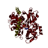
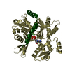
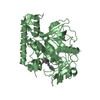
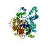
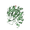
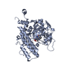
| Title | The complex between the eight-bladed symmetrical designer protein Tako8 and the silicotungstic acid Keggin (STA) | ||||||
 Components Components | Tako8 | ||||||
 Keywords Keywords | DE NOVO PROTEIN / Designer protein / polyoxometalate / POM / STA / hybrid / framework | ||||||
| Function / homology | Keggin (STA) Function and homology information Function and homology information | ||||||
| Biological species | synthetic construct (others) | ||||||
| Method |  X-RAY DIFFRACTION / SYNCHROTRON / MOLECULAR REPLACEMENT / Resolution: 2.3 Å X-RAY DIFFRACTION / SYNCHROTRON / MOLECULAR REPLACEMENT / Resolution: 2.3 Å | ||||||
 Authors Authors | Vandebroek, L. / Noguchi, H. / Parac-Vogt, T.N. / Van Meervelt, L. / Voet, A.R.D. | ||||||
 Citation Citation | Journal: Cryst.Growth Des. / Year: 2021 Title: Shape and Size Complementarity-Induced Formation of Supramolecular Protein Assemblies with Metal-Oxo Clusters Authors: Vandebroek, L. / Noguchi, H. / Kamata, K. / Tame, J.R.H. / Parac-Vogt, T.N. / Voet, A.R.D. | ||||||
| History |
|
- Structure visualization
| Structure viewer | Molecule: MolmilJmol/JSmol |
|---|
- Downloads & links
-Download
| PDBx/mmCIF format | 6y7o.cif.gz | 217 KB | Display | PDBx/mmCIF format |
|---|---|---|---|---|
| PDB format | pdb6y7o.ent.gz | 180.5 KB | Display | PDB format |
| PDBx/mmJSON format | 6y7o.json.gz | Tree view | PDBx/mmJSON format | |
| Others |  Other downloads Other downloads |
-Validation report
| Arichive directory | https://data.pdbj.org/pub/pdb/validation_reports/y7/6y7oftp://data.pdbj.org/pub/pdb/validation_reports/y7/6y7o | HTTPS FTP |
|---|
-Related structure data
| Related structure data |  6y7nC  6y7pC  6g6mS S: Starting model for refinement C: citing same article ( |
|---|---|
| Similar structure data |


-Links
 PDBj
PDBj
- Assembly
| Deposited unit | 
| |||||||||
|---|---|---|---|---|---|---|---|---|---|---|
| 1 | 
| |||||||||
| 2 | 
| |||||||||
| 3 | 
| |||||||||
| Unit cell |
| |||||||||
| Components on special symmetry positions |
|
-Components
| #1: Protein | Mass: 34981.203 Da / Num. of mol.: 3 Source method: isolated from a genetically manipulated source Source: (gene. exp.) synthetic construct (others) / Production host:  #2: Chemical | ChemComp-SIW /   Mass: 2874.142 Da / Num. of mol.: 14 / Source method: obtained synthetically / Formula: O40SiW12 / Feature type: SUBJECT OF INVESTIGATION Mass: 2874.142 Da / Num. of mol.: 14 / Source method: obtained synthetically / Formula: O40SiW12 / Feature type: SUBJECT OF INVESTIGATION#3: Water | ChemComp-HOH / |  Mass: 18.015 Da / Num. of mol.: 64 / Source method: isolated from a natural source / Formula: H2O Mass: 18.015 Da / Num. of mol.: 64 / Source method: isolated from a natural source / Formula: H2OHas ligand of interest | Y | Has protein modification | N | |
|---|
-Experimental details
-Experiment
| Experiment | Method: X-RAY DIFFRACTION / Number of used crystals: 1 |
|---|
- Sample preparation
| Crystal | Density Matthews: 2.01 Å3/Da / Density % sol: 38.72 % |
|---|---|
| Crystal grow | Temperature: 293 K / Method: vapor diffusion / pH: 4.5 / Details: 0.1M sodium acetate pH 4.5, 3.1M sodium chloride |
-Data collection
| Diffraction | Mean temperature: 100 K / Serial crystal experiment: N |
|---|---|
| Diffraction source | Source: SYNCHROTRON / Site: Diamond  / Beamline: I04-1 / Wavelength: 0.97951 Å / Beamline: I04-1 / Wavelength: 0.97951 Å |
| Detector | Type: DECTRIS PILATUS 6M / Detector: PIXEL / Date: Oct 26, 2017 |
| Radiation | Protocol: SINGLE WAVELENGTH / Monochromatic (M) / Laue (L): M / Scattering type: x-ray |
| Radiation wavelength | Wavelength: 0.97951 Å / Relative weight: 1 |
| Reflection | Resolution: 2.3→47.28 Å / Num. obs: 36727 / % possible obs: 100 % / Redundancy: 9.8 % / CC1/2: 0.989 / Rmerge(I) obs: 0.179 / Rpim(I) all: 0.09 / Rrim(I) all: 0.201 / Net I/σ(I): 8.3 |
| Reflection shell | Resolution: 2.3→2.38 Å / Rmerge(I) obs: 1.33 / Mean I/σ(I) obs: 2.5 / Num. unique obs: 3590 / CC1/2: 0.762 / Rpim(I) all: 0.679 |
- Processing
| Software |
| |||||||||||||||||||||||||||||||||||||||||||||||||||||||||||||||||||||||||||
|---|---|---|---|---|---|---|---|---|---|---|---|---|---|---|---|---|---|---|---|---|---|---|---|---|---|---|---|---|---|---|---|---|---|---|---|---|---|---|---|---|---|---|---|---|---|---|---|---|---|---|---|---|---|---|---|---|---|---|---|---|---|---|---|---|---|---|---|---|---|---|---|---|---|---|---|---|
| Refinement | Method to determine structure: MOLECULAR REPLACEMENT Starting model: 6g6m Resolution: 2.3→47.2 Å / SU ML: 0.38 / Cross valid method: THROUGHOUT / σ(F): 1.34 / Phase error: 34.49
| |||||||||||||||||||||||||||||||||||||||||||||||||||||||||||||||||||||||||||
| Solvent computation | Shrinkage radii: 0.9 Å / VDW probe radii: 1.11 Å | |||||||||||||||||||||||||||||||||||||||||||||||||||||||||||||||||||||||||||
| Displacement parameters | Biso max: 76.18 Å2 / Biso mean: 38.6316 Å2 / Biso min: 20.81 Å2 | |||||||||||||||||||||||||||||||||||||||||||||||||||||||||||||||||||||||||||
| Refinement step | Cycle: final / Resolution: 2.3→47.2 Å
| |||||||||||||||||||||||||||||||||||||||||||||||||||||||||||||||||||||||||||
| LS refinement shell | Refine-ID: X-RAY DIFFRACTION / Rfactor Rfree error: 0 / % reflection obs: 100 %
|
