 Movie
Movie Controller


+ Open data

- Basic information
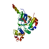
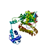
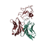
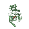
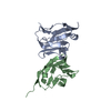
| Entry | Database: PDB / ID: 6y6r | |||||||||
|---|---|---|---|---|---|---|---|---|---|---|
| Title | Crystal structure of MINDY1 T335D mutant | |||||||||
 Components Components | Ubiquitin carboxyl-terminal hydrolase MINDY-1 | |||||||||
 Keywords Keywords | HYDROLASE / CYSTEINE PROTEASE / ISOPEPTIDASE AND UBIQUITIN BINDING | |||||||||
| Function / homology |  Function and homology information Function and homology informationcysteine-type carboxypeptidase activity / K48-linked deubiquitinase activity / K48-linked polyubiquitin modification-dependent protein binding / cell periphery / ubiquitinyl hydrolase 1 / cysteine-type deubiquitinase activity / nuclear body / proteolysis / nucleoplasm Similarity search - Function | |||||||||
| Biological species |  Homo sapiens (human) Homo sapiens (human) | |||||||||
| Method |  X-RAY DIFFRACTION / SYNCHROTRON / MOLECULAR REPLACEMENT / molecular replacement / Resolution: 3.32 Å X-RAY DIFFRACTION / SYNCHROTRON / MOLECULAR REPLACEMENT / molecular replacement / Resolution: 3.32 Å | |||||||||
 Authors Authors | Abdul Rehman, S.A. / Kulathu, Y. | |||||||||
| Funding support |  United Kingdom, 2items United Kingdom, 2items
| |||||||||
 Citation Citation | Journal: Mol.Cell / Year: 2021 Title: Mechanism of activation and regulation of deubiquitinase activity in MINDY1 and MINDY2. Authors: Abdul Rehman, S.A. / Armstrong, L.A. / Lange, S.M. / Kristariyanto, Y.A. / Grawert, T.W. / Knebel, A. / Svergun, D.I. / Kulathu, Y. | |||||||||
| History |
|
- Structure visualization
| Structure viewer | Molecule: MolmilJmol/JSmol |
|---|
- Downloads & links
-Download
| PDBx/mmCIF format | 6y6r.cif.gz | 64.9 KB | Display | PDBx/mmCIF format |
|---|---|---|---|---|
| PDB format | pdb6y6r.ent.gz | 45.7 KB | Display | PDB format |
| PDBx/mmJSON format | 6y6r.json.gz | Tree view | PDBx/mmJSON format | |
| Others |  Other downloads Other downloads |
-Validation report
| Arichive directory | https://data.pdbj.org/pub/pdb/validation_reports/y6/6y6rftp://data.pdbj.org/pub/pdb/validation_reports/y6/6y6r | HTTPS FTP |
|---|
-Related structure data
| Related structure data |  6tuvC 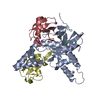 6txbC 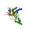 6yjgC 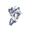 6z49C 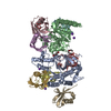 6z7vC  6z90C  7npiC 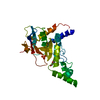 5jknS S: Starting model for refinement C: citing same article ( |
|---|---|
| Similar structure data |


-Links
 PDBj
PDBj- Assembly
| Deposited unit | 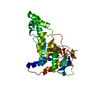
| ||||||||
|---|---|---|---|---|---|---|---|---|---|
| 1 |
| ||||||||
| Unit cell |
|
-Components
| #1: Protein | Mass: 32052.201 Da / Num. of mol.: 1 / Mutation: T335D Source method: isolated from a genetically manipulated source Source: (gene. exp.) Homo sapiens (human) / Gene: MINDY1, FAM63A, KIAA1390 / Production host:  |
|---|
-Experimental details
-Experiment
| Experiment | Method: X-RAY DIFFRACTION / Number of used crystals: 1 |
|---|
- Sample preparation
| Crystal | Density Matthews: 6.38 Å3/Da / Density % sol: 80.71 % |
|---|---|
| Crystal grow | Temperature: 293.15 K / Method: vapor diffusion, sitting drop / pH: 7.5 Details: 0.1 M HEPES sodium pH 7.5, 0.8 M Potassium sodium tartrate tetrahydrate |
-Data collection
| Diffraction | Mean temperature: 100 K / Serial crystal experiment: N | ||||||||||||||||||||||||||||||
|---|---|---|---|---|---|---|---|---|---|---|---|---|---|---|---|---|---|---|---|---|---|---|---|---|---|---|---|---|---|---|---|
| Diffraction source | Source: SYNCHROTRON / Site: Diamond / Beamline: I03 / Wavelength: 0.97628 Å | ||||||||||||||||||||||||||||||
| Detector | Type: DECTRIS PILATUS 6M / Detector: PIXEL / Date: Jan 18, 2019 | ||||||||||||||||||||||||||||||
| Radiation | Protocol: SINGLE WAVELENGTH / Monochromatic (M) / Laue (L): M / Scattering type: x-ray | ||||||||||||||||||||||||||||||
| Radiation wavelength | Wavelength: 0.97628 Å / Relative weight: 1 | ||||||||||||||||||||||||||||||
| Reflection | Resolution: 3.32→85.28 Å / Num. obs: 12828 / % possible obs: 99.4 % / Redundancy: 5.5 % / CC1/2: 0.999 / Rmerge(I) obs: 0.063 / Rpim(I) all: 0.03 / Rrim(I) all: 0.07 / Net I/σ(I): 13.2 / Num. measured all: 70094 | ||||||||||||||||||||||||||||||
| Reflection shell | Diffraction-ID: 1
|
-Phasing
| Phasing | Method: molecular replacement |
|---|
- Processing
| Software |
| ||||||||||||||||||||||||||||||||||||||||||||||||||||||||||||
|---|---|---|---|---|---|---|---|---|---|---|---|---|---|---|---|---|---|---|---|---|---|---|---|---|---|---|---|---|---|---|---|---|---|---|---|---|---|---|---|---|---|---|---|---|---|---|---|---|---|---|---|---|---|---|---|---|---|---|---|---|---|
| Refinement | Method to determine structure: MOLECULAR REPLACEMENT Starting model: 5JKN Resolution: 3.32→85.21 Å / Cor.coef. Fo:Fc: 0.952 / Cor.coef. Fo:Fc free: 0.937 / SU B: 21.483 / SU ML: 0.299 / Cross valid method: THROUGHOUT / σ(F): 0 / ESU R: 0.464 / ESU R Free: 0.333 / Stereochemistry target values: MAXIMUM LIKELIHOOD Details: HYDROGENS HAVE BEEN ADDED IN THE RIDING POSITIONS U VALUES : REFINED INDIVIDUALLY
| ||||||||||||||||||||||||||||||||||||||||||||||||||||||||||||
| Solvent computation | Ion probe radii: 0.8 Å / Shrinkage radii: 0.8 Å / VDW probe radii: 1.2 Å / Solvent model: MASK | ||||||||||||||||||||||||||||||||||||||||||||||||||||||||||||
| Displacement parameters | Biso max: 346.61 Å2 / Biso mean: 150.707 Å2 / Biso min: 30 Å2
| ||||||||||||||||||||||||||||||||||||||||||||||||||||||||||||
| Refinement step | Cycle: final / Resolution: 3.32→85.21 Å
| ||||||||||||||||||||||||||||||||||||||||||||||||||||||||||||
| Refine LS restraints |
| ||||||||||||||||||||||||||||||||||||||||||||||||||||||||||||
| LS refinement shell | Resolution: 3.32→3.401 Å / Rfactor Rfree error: 0
|
