 Movie
Movie Controller

[English] 日本語
 Yorodumi
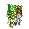
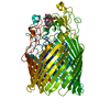
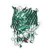
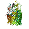
- PDB-6y47: Crystal structure of the ferric enterobactin receptor (PfeA) in c... -
+ Open data

- Basic information
| Entry | Database: PDB / ID: 6y47 | ||||||
|---|---|---|---|---|---|---|---|
| Title | Crystal structure of the ferric enterobactin receptor (PfeA) in complex with BCV-L5 | ||||||
 Components Components | Ferric enterobactin receptor | ||||||
 Keywords Keywords | MEMBRANE PROTEIN / TONB dependent transporter / siderophore | ||||||
| Function / homology |  Function and homology information Function and homology informationcolicin transmembrane transporter activity / siderophore transmembrane transport / siderophore uptake transmembrane transporter activity / siderophore transport / enterobactin transport / enterobactin transmembrane transporter activity / outer membrane / enterobactin binding / cell outer membrane / signaling receptor activity Similarity search - Function | ||||||
| Biological species |  Pseudomonas aeruginosa PAO1 (bacteria) Pseudomonas aeruginosa PAO1 (bacteria) | ||||||
| Method |  X-RAY DIFFRACTION / SYNCHROTRON / MOLECULAR REPLACEMENT / Resolution: 3.04 Å X-RAY DIFFRACTION / SYNCHROTRON / MOLECULAR REPLACEMENT / Resolution: 3.04 Å | ||||||
 Authors Authors | Naismith, J.H. / Moynie, L.M. | ||||||
| Funding support |  United Kingdom, 1items United Kingdom, 1items
| ||||||
 Citation Citation | Journal: Acs Infect Dis. / Year: 2022 Title: Hijacking of the Enterobactin Pathway by a Synthetic Catechol Vector Designed for Oxazolidinone Antibiotic Delivery in Pseudomonas aeruginosa. Authors: Moynie, L. / Hoegy, F. / Milenkovic, S. / Munier, M. / Paulen, A. / Gasser, V. / Faucon, A.L. / Zill, N. / Naismith, J.H. / Ceccarelli, M. / Schalk, I.J. / Mislin, G.L.A. | ||||||
| History |
|
- Structure visualization
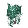
| Structure viewer | Molecule: MolmilJmol/JSmol |
|---|
- Downloads & links
-Download
| PDBx/mmCIF format | 6y47.cif.gz | 346.3 KB | Display | PDBx/mmCIF format |
|---|---|---|---|---|
| PDB format | pdb6y47.ent.gz | 237.9 KB | Display | PDB format |
| PDBx/mmJSON format | 6y47.json.gz | Tree view | PDBx/mmJSON format | |
| Others |  Other downloads Other downloads |
-Validation report
| Arichive directory | https://data.pdbj.org/pub/pdb/validation_reports/y4/6y47ftp://data.pdbj.org/pub/pdb/validation_reports/y4/6y47 | HTTPS FTP |
|---|
-Related structure data
| Related structure data | 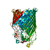 5nc3C 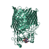 6yy5C  6z2nC  6z33C  7obwC 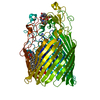 6q5eS S: Starting model for refinement C: citing same article ( |
|---|---|
| Similar structure data |


-Links
 PDBj
PDBj- Assembly
| Deposited unit | 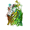
| ||||||||||||
|---|---|---|---|---|---|---|---|---|---|---|---|---|---|
| 1 |
| ||||||||||||
| Unit cell |
|
-Components
| #1: Protein | Mass: 78744.453 Da / Num. of mol.: 1 Source method: isolated from a genetically manipulated source Source: (gene. exp.) Pseudomonas aeruginosa PAO1 (bacteria) / Gene: pfeA, PA2688 / Production host: |
|---|---|
| #2: Chemical | ChemComp-OWT / ~{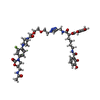  Mass: 962.976 Da / Num. of mol.: 1 / Source method: obtained synthetically / Formula: C45H55FN10O13 / Feature type: SUBJECT OF INVESTIGATION Mass: 962.976 Da / Num. of mol.: 1 / Source method: obtained synthetically / Formula: C45H55FN10O13 / Feature type: SUBJECT OF INVESTIGATION |
| #3: Chemical | ChemComp-EDO /   Mass: 62.068 Da / Num. of mol.: 1 / Source method: obtained synthetically / Formula: C2H6O2 Mass: 62.068 Da / Num. of mol.: 1 / Source method: obtained synthetically / Formula: C2H6O2 |
| #4: Chemical | ChemComp-FE /   Mass: 55.845 Da / Num. of mol.: 1 / Source method: obtained synthetically / Formula: Fe Mass: 55.845 Da / Num. of mol.: 1 / Source method: obtained synthetically / Formula: Fe |
| Has ligand of interest | Y |
| Has protein modification | Y |
-Experimental details
-Experiment
| Experiment | Method: X-RAY DIFFRACTION / Number of used crystals: 1 |
|---|
- Sample preparation
| Crystal | Density Matthews: 3.32 Å3/Da / Density % sol: 63 % |
|---|---|
| Crystal grow | Temperature: 294 K / Method: vapor diffusion / Details: PEG 8000 ADA Magnesium acetate |
-Data collection
| Diffraction | Mean temperature: 100 K / Serial crystal experiment: N |
|---|---|
| Diffraction source | Source: SYNCHROTRON / Site: Diamond / Beamline: I04-1 / Wavelength: 0.91587 Å |
| Detector | Type: DECTRIS PILATUS 6M-F / Detector: PIXEL / Date: Apr 15, 2019 |
| Radiation | Protocol: SINGLE WAVELENGTH / Monochromatic (M) / Laue (L): M / Scattering type: x-ray |
| Radiation wavelength | Wavelength: 0.91587 Å / Relative weight: 1 |
| Reflection | Resolution: 3.037→156.95 Å / Num. obs: 20933 / % possible obs: 100 % / Redundancy: 7.1 % / Biso Wilson estimate: 116.04 Å2 / CC1/2: 0.999 / Rmerge(I) obs: 0.073 / Net I/σ(I): 11.3 |
| Reflection shell | Resolution: 3.037→3.09 Å / Mean I/σ(I) obs: 1 / Num. unique obs: 982 / CC1/2: 0.53 |
- Processing
| Software |
| ||||||||||||||||||||||||||||||||||||||||||||||||||||||||
|---|---|---|---|---|---|---|---|---|---|---|---|---|---|---|---|---|---|---|---|---|---|---|---|---|---|---|---|---|---|---|---|---|---|---|---|---|---|---|---|---|---|---|---|---|---|---|---|---|---|---|---|---|---|---|---|---|---|
| Refinement | Method to determine structure: MOLECULAR REPLACEMENT Starting model: 6q5E Resolution: 3.04→69.54 Å / SU ML: 0.5766 / Cross valid method: FREE R-VALUE / σ(F): 1.34 / Phase error: 31.0801 / Stereochemistry target values: CDL v1.2
| ||||||||||||||||||||||||||||||||||||||||||||||||||||||||
| Solvent computation | Shrinkage radii: 0.9 Å / VDW probe radii: 1.11 Å / Solvent model: FLAT BULK SOLVENT MODEL | ||||||||||||||||||||||||||||||||||||||||||||||||||||||||
| Displacement parameters | Biso mean: 125.25 Å2 | ||||||||||||||||||||||||||||||||||||||||||||||||||||||||
| Refinement step | Cycle: LAST / Resolution: 3.04→69.54 Å
| ||||||||||||||||||||||||||||||||||||||||||||||||||||||||
| Refine LS restraints |
| ||||||||||||||||||||||||||||||||||||||||||||||||||||||||
| LS refinement shell |
| ||||||||||||||||||||||||||||||||||||||||||||||||||||||||
| Refinement TLS params. | Method: refined / Origin x: -83.547387746 Å / Origin y: 27.2777616146 Å / Origin z: 95.670816 Å
| ||||||||||||||||||||||||||||||||||||||||||||||||||||||||
| Refinement TLS group | Selection details: (chain A and resseq 24:901) |
