 Movie
Movie Controller

[English] 日本語
 Yorodumi
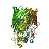
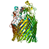
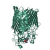
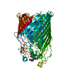
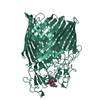
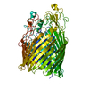
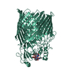
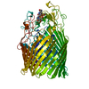
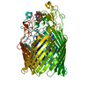
- PDB-5nr2: Crystal structure of the ferric enterobactin receptor (PfeA) from... -
+ Open data

- Basic information
| Entry | Database: PDB / ID: 5nr2 | ||||||
|---|---|---|---|---|---|---|---|
| Title | Crystal structure of the ferric enterobactin receptor (PfeA) from Pseudomonas aeruginosa in complex with azotochelin | ||||||
 Components Components | Ferric enterobactin receptor | ||||||
 Keywords Keywords | MEMBRANE PROTEIN / PfeA / PA2688 / outer membrane receptor / azotochelin | ||||||
| Function / homology |  Function and homology information Function and homology informationcolicin transmembrane transporter activity / siderophore transmembrane transport / siderophore uptake transmembrane transporter activity / siderophore transport / enterobactin transport / enterobactin transmembrane transporter activity / outer membrane / enterobactin binding / cell outer membrane / signaling receptor activity Similarity search - Function | ||||||
| Biological species |   Pseudomonas aeruginosa (bacteria) Pseudomonas aeruginosa (bacteria) | ||||||
| Method |  X-RAY DIFFRACTION / SYNCHROTRON / MOLECULAR REPLACEMENT / Resolution: 2.78 Å X-RAY DIFFRACTION / SYNCHROTRON / MOLECULAR REPLACEMENT / Resolution: 2.78 Å | ||||||
 Authors Authors | Moynie, L. / Naismith, J.H. | ||||||
 Citation Citation | Journal: Nat Commun / Year: 2019 Title: The complex of ferric-enterobactin with its transporter from Pseudomonas aeruginosa suggests a two-site model. Authors: Moynie, L. / Milenkovic, S. / Mislin, G.L.A. / Gasser, V. / Malloci, G. / Baco, E. / McCaughan, R.P. / Page, M.G.P. / Schalk, I.J. / Ceccarelli, M. / Naismith, J.H. | ||||||
| History |
|
- Structure visualization
| Structure viewer | Molecule: MolmilJmol/JSmol |
|---|
- Downloads & links
-Download
| PDBx/mmCIF format | 5nr2.cif.gz | 289.9 KB | Display | PDBx/mmCIF format |
|---|---|---|---|---|
| PDB format | pdb5nr2.ent.gz | 236 KB | Display | PDB format |
| PDBx/mmJSON format | 5nr2.json.gz | Tree view | PDBx/mmJSON format | |
| Others |  Other downloads Other downloads |
-Validation report
| Arichive directory | https://data.pdbj.org/pub/pdb/validation_reports/nr/5nr2ftp://data.pdbj.org/pub/pdb/validation_reports/nr/5nr2 | HTTPS FTP |
|---|
-Related structure data
| Related structure data | 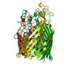 5m9bC 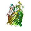 5mzsC  5nc4C  5outC  6i2jC 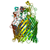 6q5eC  6r1fC 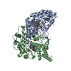 1m9kS S: Starting model for refinement C: citing same article ( |
|---|---|
| Similar structure data |

-Links
 PDBj
PDBj- Assembly
| Deposited unit | 
| ||||||||
|---|---|---|---|---|---|---|---|---|---|
| 1 |
| ||||||||
| Unit cell |
|
-Components
| #1: Protein | Mass: 78744.453 Da / Num. of mol.: 1 Source method: isolated from a genetically manipulated source Source: (gene. exp.) Pseudomonas aeruginosa (bacteria) / Strain: PAO1 / Gene: pfeA, PA2688 / Production host: |
|---|---|
| #2: Chemical | ChemComp-FE /   Mass: 55.845 Da / Num. of mol.: 1 / Source method: obtained synthetically / Formula: Fe Mass: 55.845 Da / Num. of mol.: 1 / Source method: obtained synthetically / Formula: Fe |
| #3: Chemical | ChemComp-95B / 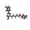  Mass: 418.397 Da / Num. of mol.: 1 / Source method: obtained synthetically / Formula: C20H22N2O8 Mass: 418.397 Da / Num. of mol.: 1 / Source method: obtained synthetically / Formula: C20H22N2O8 |
| #4: Chemical | ChemComp-EDO /   Mass: 62.068 Da / Num. of mol.: 1 / Source method: obtained synthetically / Formula: C2H6O2 Mass: 62.068 Da / Num. of mol.: 1 / Source method: obtained synthetically / Formula: C2H6O2 |
| Has protein modification | Y |
-Experimental details
-Experiment
| Experiment | Method: X-RAY DIFFRACTION / Number of used crystals: 1 |
|---|
- Sample preparation
| Crystal | Density Matthews: 3.55 Å3/Da / Density % sol: 65.39 % |
|---|---|
| Crystal grow | Temperature: 293 K / Method: vapor diffusion / pH: 6.5 / Details: PEG 8000, ADA, Magnesium acetate |
-Data collection
| Diffraction | Mean temperature: 100 K |
|---|---|
| Diffraction source | Source: SYNCHROTRON / Site: Diamond  / Beamline: I03 / Wavelength: 0.9763 Å / Beamline: I03 / Wavelength: 0.9763 Å |
| Detector | Type: DECTRIS PILATUS3 S 6M / Detector: PIXEL / Date: Aug 6, 2016 |
| Radiation | Protocol: SINGLE WAVELENGTH / Monochromatic (M) / Laue (L): M / Scattering type: x-ray |
| Radiation wavelength | Wavelength: 0.9763 Å / Relative weight: 1 |
| Reflection | Resolution: 2.78→86.64 Å / Num. obs: 28035 / % possible obs: 99.9 % / Redundancy: 7.3 % / CC1/2: 0.999 / Rmerge(I) obs: 0.048 / Net I/σ(I): 20.8 |
| Reflection shell | Resolution: 2.78→2.85 Å / Redundancy: 7.3 % / Rmerge(I) obs: 1.057 / Mean I/σ(I) obs: 1.8 / Num. unique obs: 2036 / CC1/2: 0.677 / % possible all: 99.8 |
- Processing
| Software |
| ||||||||||||||||||||||||||||||||||||||||||||||||||||||||||||||||||||||||||||||||||||||||||||||||||||||||||||||||||||||||||||||||||||||||||||||||||||||||||||||||||||||||||||||||||||||
|---|---|---|---|---|---|---|---|---|---|---|---|---|---|---|---|---|---|---|---|---|---|---|---|---|---|---|---|---|---|---|---|---|---|---|---|---|---|---|---|---|---|---|---|---|---|---|---|---|---|---|---|---|---|---|---|---|---|---|---|---|---|---|---|---|---|---|---|---|---|---|---|---|---|---|---|---|---|---|---|---|---|---|---|---|---|---|---|---|---|---|---|---|---|---|---|---|---|---|---|---|---|---|---|---|---|---|---|---|---|---|---|---|---|---|---|---|---|---|---|---|---|---|---|---|---|---|---|---|---|---|---|---|---|---|---|---|---|---|---|---|---|---|---|---|---|---|---|---|---|---|---|---|---|---|---|---|---|---|---|---|---|---|---|---|---|---|---|---|---|---|---|---|---|---|---|---|---|---|---|---|---|---|---|
| Refinement | Method to determine structure: MOLECULAR REPLACEMENT Starting model: 1m9k Resolution: 2.78→86.64 Å / Cor.coef. Fo:Fc: 0.876 / Cor.coef. Fo:Fc free: 0.922 / SU B: 34.384 / SU ML: 0.3 / Cross valid method: THROUGHOUT / ESU R: 0.655 / ESU R Free: 0.331 / Details: HYDROGENS HAVE BEEN ADDED IN THE RIDING POSITIONS
| ||||||||||||||||||||||||||||||||||||||||||||||||||||||||||||||||||||||||||||||||||||||||||||||||||||||||||||||||||||||||||||||||||||||||||||||||||||||||||||||||||||||||||||||||||||||
| Solvent computation | Ion probe radii: 0.8 Å / Shrinkage radii: 0.8 Å / VDW probe radii: 1.2 Å | ||||||||||||||||||||||||||||||||||||||||||||||||||||||||||||||||||||||||||||||||||||||||||||||||||||||||||||||||||||||||||||||||||||||||||||||||||||||||||||||||||||||||||||||||||||||
| Displacement parameters | Biso mean: 107.054 Å2
| ||||||||||||||||||||||||||||||||||||||||||||||||||||||||||||||||||||||||||||||||||||||||||||||||||||||||||||||||||||||||||||||||||||||||||||||||||||||||||||||||||||||||||||||||||||||
| Refinement step | Cycle: 1 / Resolution: 2.78→86.64 Å
| ||||||||||||||||||||||||||||||||||||||||||||||||||||||||||||||||||||||||||||||||||||||||||||||||||||||||||||||||||||||||||||||||||||||||||||||||||||||||||||||||||||||||||||||||||||||
| Refine LS restraints |
|