 Movie
Movie Controller

[English] 日本語
 Yorodumi
- PDB-6y3z: Crystal structure of the Pby1 ATP-grasp enzyme bound to the S. ce... -
+ Open data

- Basic information
| Entry | Database: PDB / ID: 6y3z | ||||||||||||
|---|---|---|---|---|---|---|---|---|---|---|---|---|---|
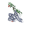
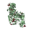
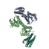
| Title | Crystal structure of the Pby1 ATP-grasp enzyme bound to the S. cerevisiae mRNA decapping complex (Dcp1-Dcp2-Edc3) | ||||||||||||
 Components Components |
| ||||||||||||
 Keywords Keywords | HYDROLASE / RNA decay enzyme | ||||||||||||
| Function / homology |  Function and homology information Function and homology informationRNA decapping complex / tubulin-tyrosine ligase / Butyrate Response Factor 1 (BRF1) binds and destabilizes mRNA / Tristetraprolin (TTP, ZFP36) binds and destabilizes mRNA / mRNA decay by 5' to 3' exoribonuclease / deadenylation-independent decapping of nuclear-transcribed mRNA / 5'-(N7-methylguanosine 5'-triphospho)-[mRNA] hydrolase / 5'-(N(7)-methylguanosine 5'-triphospho)-[mRNA] hydrolase activity / deadenylation-dependent decapping of nuclear-transcribed mRNA / cytoplasmic side of membrane ...RNA decapping complex / tubulin-tyrosine ligase / Butyrate Response Factor 1 (BRF1) binds and destabilizes mRNA / Tristetraprolin (TTP, ZFP36) binds and destabilizes mRNA / mRNA decay by 5' to 3' exoribonuclease / deadenylation-independent decapping of nuclear-transcribed mRNA / 5'-(N7-methylguanosine 5'-triphospho)-[mRNA] hydrolase / 5'-(N(7)-methylguanosine 5'-triphospho)-[mRNA] hydrolase activity / deadenylation-dependent decapping of nuclear-transcribed mRNA / cytoplasmic side of membrane / tubulin-tyrosine ligase activity / nuclear-transcribed mRNA catabolic process, nonsense-mediated decay / P-body assembly / positive regulation of nuclear-transcribed mRNA catabolic process, deadenylation-dependent decay / positive regulation of transcription initiation by RNA polymerase II / cellular response to glucose starvation / stress granule assembly / protein modification process / P-body / enzyme activator activity / mRNA processing / manganese ion binding / hydrolase activity / mRNA binding / chromatin binding / ATP binding / nucleus / cytoplasm Similarity search - Function | ||||||||||||
| Biological species |  | ||||||||||||
| Method |  X-RAY DIFFRACTION / SYNCHROTRON / MOLECULAR REPLACEMENT / Resolution: 3.49 Å X-RAY DIFFRACTION / SYNCHROTRON / MOLECULAR REPLACEMENT / Resolution: 3.49 Å | ||||||||||||
 Authors Authors | Graille, M. | ||||||||||||
| Funding support |  France, 3items France, 3items
| ||||||||||||
 Citation Citation | Journal: Nucleic Acids Res. / Year: 2020 Title: Pby1 is a direct partner of the Dcp2 decapping enzyme. Authors: Charenton, C. / Gaudon-Plesse, C. / Back, R. / Ulryck, N. / Cosson, L. / Seraphin, B. / Graille, M. | ||||||||||||
| History |
|
- Structure visualization
| Structure viewer | Molecule: MolmilJmol/JSmol |
|---|
- Downloads & links
-Download
| PDBx/mmCIF format | 6y3z.cif.gz | 304 KB | Display | PDBx/mmCIF format |
|---|---|---|---|---|
| PDB format | pdb6y3z.ent.gz | 244.1 KB | Display | PDB format |
| PDBx/mmJSON format | 6y3z.json.gz | Tree view | PDBx/mmJSON format | |
| Others |  Other downloads Other downloads |
-Validation report
| Arichive directory | https://data.pdbj.org/pub/pdb/validation_reports/y3/6y3zftp://data.pdbj.org/pub/pdb/validation_reports/y3/6y3z | HTTPS FTP |
|---|
-Related structure data
| Related structure data |  6y3pSC 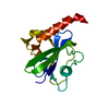 4k6eS  5lopS S: Starting model for refinement C: citing same article ( |
|---|---|
| Similar structure data |


-Links
 PDBj
PDBj


- Assembly
| Deposited unit | 
| ||||||||
|---|---|---|---|---|---|---|---|---|---|
| 1 |
| ||||||||
| Unit cell |
|
-Components
| #1: Protein | Mass: 32946.887 Da / Num. of mol.: 1 Source method: isolated from a genetically manipulated source Source: (gene. exp.) Production host:  References: UniProt: P53550, 5'-(N7-methylguanosine 5'-triphospho)-[mRNA] hydrolase |
|---|---|
| #2: Protein | Mass: 26705.814 Da / Num. of mol.: 1 Source method: isolated from a genetically manipulated source Source: (gene. exp.) Production host: References: UniProt: Q12517 |
| #3: Protein | Mass: 7448.622 Da / Num. of mol.: 1 Source method: isolated from a genetically manipulated source Source: (gene. exp.) Production host: References: UniProt: P39998 |
| #4: Protein | Mass: 49895.680 Da / Num. of mol.: 1 Source method: isolated from a genetically manipulated source Source: (gene. exp.) Production host: References: UniProt: P38254, tubulin-tyrosine ligase |
| #5: Chemical | ChemComp-MG /   Mass: 24.305 Da / Num. of mol.: 1 / Source method: obtained synthetically / Formula: Mg / Feature type: SUBJECT OF INVESTIGATION Mass: 24.305 Da / Num. of mol.: 1 / Source method: obtained synthetically / Formula: Mg / Feature type: SUBJECT OF INVESTIGATION |
| Has ligand of interest | Y |
-Experimental details
-Experiment
| Experiment | Method: X-RAY DIFFRACTION / Number of used crystals: 1 |
|---|
- Sample preparation
| Crystal | Density Matthews: 2.68 Å3/Da / Density % sol: 54.07 % |
|---|---|
| Crystal grow | Temperature: 277 K / Method: vapor diffusion, hanging drop Details: 0.05 M calcium acetate; 0.1 M sodium cacodylate pH6; 25% MPD |
-Data collection
| Diffraction | Mean temperature: 100 K / Serial crystal experiment: N |
|---|---|
| Diffraction source | Source: SYNCHROTRON / Site: ESRF / Beamline: ID23-1 / Wavelength: 0.97626 Å |
| Detector | Type: DECTRIS PILATUS3 S 6M / Detector: PIXEL / Date: Feb 16, 2015 |
| Radiation | Protocol: SINGLE WAVELENGTH / Monochromatic (M) / Laue (L): M / Scattering type: x-ray |
| Radiation wavelength | Wavelength: 0.97626 Å / Relative weight: 1 |
| Reflection | Resolution: 3.49→48.5 Å / Num. obs: 16546 / % possible obs: 99.5 % / Redundancy: 10.1 % / CC1/2: 0.999 / Rmerge(I) obs: 0.101 / Rpim(I) all: 0.034 / Rsym value: 0.107 / Net I/σ(I): 9.6 |
| Reflection shell | Resolution: 3.49→3.55 Å / Rmerge(I) obs: 1.409 / Mean I/σ(I) obs: 1.3 / Num. unique obs: 828 / CC1/2: 0.07 / Rpim(I) all: 0.479 / Rsym value: 1.494 |
- Processing
| Software |
| ||||||||||||||||||||||||||||||||||||||||||||||||||||||||||||||||||||||||||||||||||||||||||||||||||||||||||||
|---|---|---|---|---|---|---|---|---|---|---|---|---|---|---|---|---|---|---|---|---|---|---|---|---|---|---|---|---|---|---|---|---|---|---|---|---|---|---|---|---|---|---|---|---|---|---|---|---|---|---|---|---|---|---|---|---|---|---|---|---|---|---|---|---|---|---|---|---|---|---|---|---|---|---|---|---|---|---|---|---|---|---|---|---|---|---|---|---|---|---|---|---|---|---|---|---|---|---|---|---|---|---|---|---|---|---|---|---|---|
| Refinement | Method to determine structure: MOLECULAR REPLACEMENT Starting model: 4K6E, 6Y3P, 5LOP Resolution: 3.49→48.5 Å / Cor.coef. Fo:Fc: 0.899 / Cor.coef. Fo:Fc free: 0.876 / Rfactor Rfree error: 0 / Cross valid method: THROUGHOUT / σ(F): 0 / SU Rfree Blow DPI: 0.638
| ||||||||||||||||||||||||||||||||||||||||||||||||||||||||||||||||||||||||||||||||||||||||||||||||||||||||||||
| Displacement parameters | Biso max: 300 Å2 / Biso mean: 193.18 Å2 / Biso min: 83.52 Å2
| ||||||||||||||||||||||||||||||||||||||||||||||||||||||||||||||||||||||||||||||||||||||||||||||||||||||||||||
| Refine analyze | Luzzati coordinate error obs: 0.54 Å | ||||||||||||||||||||||||||||||||||||||||||||||||||||||||||||||||||||||||||||||||||||||||||||||||||||||||||||
| Refinement step | Cycle: final / Resolution: 3.49→48.5 Å
| ||||||||||||||||||||||||||||||||||||||||||||||||||||||||||||||||||||||||||||||||||||||||||||||||||||||||||||
| Refine LS restraints |
| ||||||||||||||||||||||||||||||||||||||||||||||||||||||||||||||||||||||||||||||||||||||||||||||||||||||||||||
| LS refinement shell | Resolution: 3.49→3.73 Å / Rfactor Rfree error: 0 / Total num. of bins used: 8
| ||||||||||||||||||||||||||||||||||||||||||||||||||||||||||||||||||||||||||||||||||||||||||||||||||||||||||||
| Refinement TLS params. | Method: refined / Refine-ID: X-RAY DIFFRACTION
| ||||||||||||||||||||||||||||||||||||||||||||||||||||||||||||||||||||||||||||||||||||||||||||||||||||||||||||
| Refinement TLS group |
|
