 Movie
Movie Controller

[English] 日本語
 Yorodumi
- PDB-6y2o: Crystal structure of the cAMP-dependent protein kinase A cocrysta... -
+ Open data

- Basic information
| Entry | Database: PDB / ID: 6y2o | ||||||
|---|---|---|---|---|---|---|---|
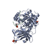
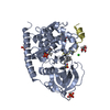
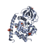
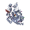
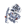
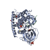
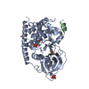
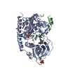
| Title | Crystal structure of the cAMP-dependent protein kinase A cocrystallized with 1,7-Naphthyridin-8-amine and PKI (5-24) | ||||||
 Components Components |
| ||||||
 Keywords Keywords | TRANSFERASE / phosphotransferase / signalling pathways / glycogen metabolism / serine/threonine kinase | ||||||
| Function / homology |  Function and homology information Function and homology informationcAMP-dependent protein kinase inhibitor activity / histone H1-4S35 kinase activity / cAMP-dependent protein kinase / regulation of protein processing / dorsal/ventral neural tube patterning / cAMP-dependent protein kinase activity / cellular response to parathyroid hormone stimulus / protein localization to lipid droplet / negative regulation of protein import into nucleus / regulation of bicellular tight junction assembly ...cAMP-dependent protein kinase inhibitor activity / histone H1-4S35 kinase activity / cAMP-dependent protein kinase / regulation of protein processing / dorsal/ventral neural tube patterning / cAMP-dependent protein kinase activity / cellular response to parathyroid hormone stimulus / protein localization to lipid droplet / negative regulation of protein import into nucleus / regulation of bicellular tight junction assembly / cAMP-dependent protein kinase complex / interleukin-2-mediated signaling pathway / sperm capacitation / regulation of osteoblast differentiation / cellular response to cold / protein kinase A regulatory subunit binding / negative regulation of glycolytic process through fructose-6-phosphate / ciliary base / protein kinase A catalytic subunit binding / intracellular potassium ion homeostasis / TORC1 signaling / mesoderm formation / plasma membrane raft / axoneme / sperm flagellum / postsynaptic modulation of chemical synaptic transmission / regulation of G2/M transition of mitotic cell cycle / negative regulation of cAMP/PKA signal transduction / negative regulation of TORC1 signaling / protein serine/threonine/tyrosine kinase activity / protein export from nucleus / cellular response to glucagon stimulus / cellular response to nutrient levels / positive regulation of gluconeogenesis / positive regulation of phagocytosis / acrosomal vesicle / regulation of proteasomal protein catabolic process / positive regulation of protein export from nucleus / neural tube closure / cellular response to glucose stimulus / negative regulation of smoothened signaling pathway / neuromuscular junction / positive regulation of cholesterol biosynthetic process / mRNA processing / positive regulation of insulin secretion / adenylate cyclase-inhibiting G protein-coupled receptor signaling pathway / manganese ion binding / cellular response to heat / adenylate cyclase-activating G protein-coupled receptor signaling pathway / postsynapse / regulation of cell cycle / nuclear speck / protein domain specific binding / protein serine kinase activity / protein serine/threonine kinase activity / centrosome / ubiquitin protein ligase binding / protein kinase binding / perinuclear region of cytoplasm / glutamatergic synapse / magnesium ion binding / negative regulation of transcription by RNA polymerase II / mitochondrion / ATP binding / nucleus / cytosol / cytoplasm Similarity search - Function | ||||||
| Biological species |   Cricetulus griseus (Chinese hamster) Cricetulus griseus (Chinese hamster) | ||||||
| Method |  X-RAY DIFFRACTION / SYNCHROTRON / MOLECULAR REPLACEMENT / Resolution: 2.01 Å X-RAY DIFFRACTION / SYNCHROTRON / MOLECULAR REPLACEMENT / Resolution: 2.01 Å | ||||||
 Authors Authors | Oebbeke, M. / Heine, A. / Klebe, G. | ||||||
 Citation Citation | Journal: To Be Published Title: Fragment based drug design - Small chemical changes of fragments effecting big changes in binding Authors: Oebbeke, M. / Wienen-Schmidt, B. / Gerber, H.-D. / Heine, A. / Klebe, G. | ||||||
| History |
|
- Structure visualization
| Structure viewer | Molecule: MolmilJmol/JSmol |
|---|
- Downloads & links
-Download
| PDBx/mmCIF format | 6y2o.cif.gz | 105.8 KB | Display | PDBx/mmCIF format |
|---|---|---|---|---|
| PDB format | pdb6y2o.ent.gz | 66.8 KB | Display | PDB format |
| PDBx/mmJSON format | 6y2o.json.gz | Tree view | PDBx/mmJSON format | |
| Others |  Other downloads Other downloads |
-Validation report
| Arichive directory | https://data.pdbj.org/pub/pdb/validation_reports/y2/6y2oftp://data.pdbj.org/pub/pdb/validation_reports/y2/6y2o | HTTPS FTP |
|---|
-Related structure data
| Related structure data |  6f14SC  6y05C  6y0bC 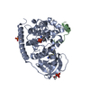 6y2uC  6y89C 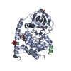 6yotC 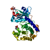 6youC  7axtC  7axvC  7axwC S: Starting model for refinement C: citing same article ( |
|---|---|
| Similar structure data |


-Links
 PDBj
PDBj


- Assembly
| Deposited unit | 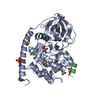
| ||||||||||||
|---|---|---|---|---|---|---|---|---|---|---|---|---|---|
| 1 |
| ||||||||||||
| Unit cell |
|
-Components
| #1: Protein | Mass: 41113.766 Da / Num. of mol.: 1 Source method: isolated from a genetically manipulated source Source: (gene. exp.) Cricetulus griseus (Chinese hamster) / Gene: PRKACA / Production host:  | ||||||||
|---|---|---|---|---|---|---|---|---|---|
| #2: Protein/peptide | Mass: 2226.411 Da / Num. of mol.: 1 / Source method: obtained synthetically / Source: (synth.) | ||||||||
| #3: Chemical | ChemComp-DMS /   Mass: 78.133 Da / Num. of mol.: 5 / Source method: obtained synthetically / Formula: C2H6OS / Comment: DMSO, precipitant*YM Mass: 78.133 Da / Num. of mol.: 5 / Source method: obtained synthetically / Formula: C2H6OS / Comment: DMSO, precipitant*YM#4: Chemical | ChemComp-O72 / |   Mass: 145.161 Da / Num. of mol.: 1 / Source method: obtained synthetically / Formula: C8H7N3 / Feature type: SUBJECT OF INVESTIGATION Mass: 145.161 Da / Num. of mol.: 1 / Source method: obtained synthetically / Formula: C8H7N3 / Feature type: SUBJECT OF INVESTIGATION#5: Water | ChemComp-HOH / |  Mass: 18.015 Da / Num. of mol.: 134 / Source method: isolated from a natural source / Formula: H2O Mass: 18.015 Da / Num. of mol.: 134 / Source method: isolated from a natural source / Formula: H2OHas ligand of interest | Y | Has protein modification | Y | |
-Experimental details
-Experiment
| Experiment | Method: X-RAY DIFFRACTION / Number of used crystals: 1 |
|---|
- Sample preparation
| Crystal | Density Matthews: 2.84 Å3/Da / Density % sol: 56.66 % |
|---|---|
| Crystal grow | Temperature: 277 K / Method: vapor diffusion, hanging drop / pH: 6.9 Details: 30 mM MES-BIS-Tris-Buffer, 1 mM dithiothreitol, 0.1 mM sodium EDTA, 75 mM LiCl, 0.2 Mega8, 14 mM 1,7-Naphthyridin-8-amine, 0.5 mM PKI (5-24) and 18-23 % methanol (v/v)0.004 mL drop volume, 1. ...Details: 30 mM MES-BIS-Tris-Buffer, 1 mM dithiothreitol, 0.1 mM sodium EDTA, 75 mM LiCl, 0.2 Mega8, 14 mM 1,7-Naphthyridin-8-amine, 0.5 mM PKI (5-24) and 18-23 % methanol (v/v)0.004 mL drop volume, 1.0 mL reservoir volume |
-Data collection
| Diffraction | Mean temperature: 100 K / Serial crystal experiment: N |
|---|---|
| Diffraction source | Source: SYNCHROTRON / Site: BESSY  / Beamline: 14.1 / Wavelength: 0.9184 Å / Beamline: 14.1 / Wavelength: 0.9184 Å |
| Detector | Type: DECTRIS PILATUS 6M / Detector: PIXEL / Date: Apr 6, 2018 |
| Radiation | Protocol: SINGLE WAVELENGTH / Monochromatic (M) / Laue (L): M / Scattering type: x-ray |
| Radiation wavelength | Wavelength: 0.9184 Å / Relative weight: 1 |
| Reflection | Resolution: 2.006→46.18 Å / Num. obs: 31850 / % possible obs: 99.5 % / Redundancy: 5.9 % / Rsym value: 0.054 / Net I/σ(I): 19.9 |
| Reflection shell | Resolution: 2.01→2.13 Å / Mean I/σ(I) obs: 3.56 / Num. unique obs: 5005 / Rsym value: 0.489 / % possible all: 98.4 |
- Processing
| Software |
| ||||||||||||||||||||||||||||||||||||||||||||||||||||||||||||||||||||||||||||||||||||
|---|---|---|---|---|---|---|---|---|---|---|---|---|---|---|---|---|---|---|---|---|---|---|---|---|---|---|---|---|---|---|---|---|---|---|---|---|---|---|---|---|---|---|---|---|---|---|---|---|---|---|---|---|---|---|---|---|---|---|---|---|---|---|---|---|---|---|---|---|---|---|---|---|---|---|---|---|---|---|---|---|---|---|---|---|---|
| Refinement | Method to determine structure: MOLECULAR REPLACEMENT Starting model: 6F14 Resolution: 2.01→46.18 Å / SU ML: 0.2256 / Cross valid method: FREE R-VALUE / σ(F): 1.37 / Phase error: 25.9412
| ||||||||||||||||||||||||||||||||||||||||||||||||||||||||||||||||||||||||||||||||||||
| Solvent computation | Shrinkage radii: 0.9 Å / VDW probe radii: 1.11 Å | ||||||||||||||||||||||||||||||||||||||||||||||||||||||||||||||||||||||||||||||||||||
| Displacement parameters | Biso mean: 37.8 Å2 | ||||||||||||||||||||||||||||||||||||||||||||||||||||||||||||||||||||||||||||||||||||
| Refinement step | Cycle: LAST / Resolution: 2.01→46.18 Å
| ||||||||||||||||||||||||||||||||||||||||||||||||||||||||||||||||||||||||||||||||||||
| Refine LS restraints |
| ||||||||||||||||||||||||||||||||||||||||||||||||||||||||||||||||||||||||||||||||||||
| LS refinement shell |
|
