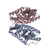
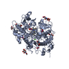
[English] 日本語
Yorodumi
- PDB-6y20: Crystal structure of Protein Scalloped (222-440) bound to Protein... -

+
Open data


ID or keywords:

Loading...

-
Basic information

Entry
Database: PDB / ID: 6y20
TitleCrystal structure of Protein Scalloped (222-440) bound to Protein Vestigial (298-337)
Components
  • (Protein scalloped) x 2
  • Protein vestigial
KeywordsTRANSCRIPTION / Complex
Function / homology
Function and homology information


dorsal/ventral lineage restriction, imaginal disc / determination of muscle attachment site / cell elongation involved in imaginal disc-derived wing morphogenesis / : / Transcriptional activation by YKI / : / wing disc proximal/distal pattern formation / haltere development / wing disc dorsal/ventral pattern formation / imaginal disc-derived leg morphogenesis ...dorsal/ventral lineage restriction, imaginal disc / determination of muscle attachment site / cell elongation involved in imaginal disc-derived wing morphogenesis / : / Transcriptional activation by YKI / : / wing disc proximal/distal pattern formation / haltere development / wing disc dorsal/ventral pattern formation / imaginal disc-derived leg morphogenesis / somatic muscle development / compound eye morphogenesis / regulation of heart morphogenesis / wing disc morphogenesis / wing disc development / imaginal disc-derived wing morphogenesis / : / sensory organ development / hippo signaling / cardiac muscle cell development / negative regulation of neuroblast proliferation / embryonic organ development / regulation of mitotic cell cycle / transcription corepressor binding / stem cell proliferation / transcription coactivator binding / RNA polymerase II transcription regulator complex / regulation of cell population proliferation / DNA replication / transcription regulator complex / transcription cis-regulatory region binding / DNA-binding transcription factor activity, RNA polymerase II-specific / cell cycle / RNA polymerase II cis-regulatory region sequence-specific DNA binding / protein domain specific binding / negative regulation of DNA-templated transcription / regulation of transcription by RNA polymerase II / positive regulation of transcription by RNA polymerase II / DNA binding / nucleoplasm / nucleus / metal ion binding
Similarity search - Function
Vestigial family / Vestigial/Tondu family / TDU repeat / Short repeats in human TONDU, fly vestigial and other proteins. / TEA/ATTS domain / Transcriptional enhancer factor, metazoa / TEA/ATTS domain superfamily / TEA/ATTS domain / TEA domain signature. / TEA domain profile. ...Vestigial family / Vestigial/Tondu family / TDU repeat / Short repeats in human TONDU, fly vestigial and other proteins. / TEA/ATTS domain / Transcriptional enhancer factor, metazoa / TEA/ATTS domain superfamily / TEA/ATTS domain / TEA domain signature. / TEA domain profile. / TEA domain / YAP binding domain / YAP binding domain
Similarity search - Domain/homology
MYRISTIC ACID / Protein scalloped / Protein vestigial
Similarity search - Component
Biological speciesDrosophila melanogaster (fruit fly)
MethodX-RAY DIFFRACTION / SYNCHROTRON / MOLECULAR REPLACEMENT / Resolution: 1.849 Å
AuthorsScheufler, C. / Villard, F. / Bokhovchuk, F.
CitationJournal: Sci Rep / Year: 2020
Title: A new perspective on the interaction between the Vg/VGLL1-3 proteins and the TEAD transcription factors.
Authors: Mesrouze, Y. / Aguilar, G. / Bokhovchuk, F. / Martin, T. / Delaunay, C. / Villard, F. / Meyerhofer, M. / Zimmermann, C. / Fontana, P. / Wille, R. / Vorherr, T. / Erdmann, D. / Furet, P. / ...Authors: Mesrouze, Y. / Aguilar, G. / Bokhovchuk, F. / Martin, T. / Delaunay, C. / Villard, F. / Meyerhofer, M. / Zimmermann, C. / Fontana, P. / Wille, R. / Vorherr, T. / Erdmann, D. / Furet, P. / Scheufler, C. / Schmelzle, T. / Affolter, M. / Chene, P.
History
DepositionFeb 14, 2020Deposition site: PDBE / Processing site: PDBE
Revision 1.0Oct 21, 2020Provider: repository / Type: Initial release
Revision 1.1Oct 28, 2020Group: Database references / Category: citation / citation_author
Item: _citation.journal_volume / _citation.page_first ..._citation.journal_volume / _citation.page_first / _citation.page_last / _citation.pdbx_database_id_PubMed / _citation.title
Revision 1.2Jan 24, 2024Group: Data collection / Database references / Refinement description
Category: chem_comp_atom / chem_comp_bond ...chem_comp_atom / chem_comp_bond / database_2 / pdbx_initial_refinement_model
Item: _database_2.pdbx_DOI / _database_2.pdbx_database_accession
Revision 2.0May 29, 2024Group: Atomic model / Data collection ...Atomic model / Data collection / Database references / Derived calculations / Polymer sequence / Source and taxonomy / Structure summary
Category: atom_site / atom_site_anisotrop ...atom_site / atom_site_anisotrop / entity / entity_poly / entity_poly_seq / entity_src_gen / pdbx_entity_nonpoly / pdbx_entity_src_syn / pdbx_nonpoly_scheme / pdbx_poly_seq_scheme / pdbx_struct_assembly_gen / struct_asym / struct_conn / struct_ref_seq_dif / struct_site / struct_site_gen
Item: _atom_site.B_iso_or_equiv / _atom_site.Cartn_x ..._atom_site.B_iso_or_equiv / _atom_site.Cartn_x / _atom_site.Cartn_y / _atom_site.Cartn_z / _atom_site.auth_asym_id / _atom_site.auth_atom_id / _atom_site.auth_comp_id / _atom_site.auth_seq_id / _atom_site.group_PDB / _atom_site.label_asym_id / _atom_site.label_atom_id / _atom_site.label_comp_id / _atom_site.label_entity_id / _atom_site.label_seq_id / _atom_site.type_symbol / _atom_site_anisotrop.U[1][1] / _atom_site_anisotrop.U[1][2] / _atom_site_anisotrop.U[1][3] / _atom_site_anisotrop.U[2][2] / _atom_site_anisotrop.U[2][3] / _atom_site_anisotrop.U[3][3] / _atom_site_anisotrop.pdbx_auth_asym_id / _atom_site_anisotrop.pdbx_auth_atom_id / _atom_site_anisotrop.pdbx_auth_comp_id / _atom_site_anisotrop.pdbx_auth_seq_id / _atom_site_anisotrop.pdbx_label_asym_id / _atom_site_anisotrop.pdbx_label_atom_id / _atom_site_anisotrop.pdbx_label_comp_id / _atom_site_anisotrop.pdbx_label_seq_id / _atom_site_anisotrop.type_symbol / _entity_poly.pdbx_seq_one_letter_code / _entity_poly.pdbx_seq_one_letter_code_can / _entity_src_gen.gene_src_common_name / _pdbx_entity_src_syn.organism_common_name / _pdbx_entity_src_syn.pdbx_end_seq_num / _pdbx_struct_assembly_gen.asym_id_list / _struct_conn.ptnr2_auth_seq_id / _struct_conn.ptnr2_label_asym_id / _struct_conn.ptnr2_label_seq_id / _struct_site.details / _struct_site.pdbx_auth_seq_id / _struct_site_gen.label_asym_id

-
Structure visualization

Structure viewerMolecule:
MolmilJmol/JSmol

Downloads & links

-
Assembly

Deposited unit
A: Protein scalloped
B: Protein scalloped
C: Protein vestigial
D: Protein vestigial
hetero molecules


Theoretical massNumber of molelcules
Total (without water)60,7765
Polymers60,5474
Non-polymers2281
Water4,594255
1


  • Idetical with deposited unit
  • defined by author&software
TypeNameSymmetry operationNumber
identity operation1_555x,y,z1
Buried area8630 Å2
ΔGint-28 kcal/mol
Surface area22280 Å2
MethodPISA
Unit cell
Length a, b, c (Å)48.23, 62.039, 156.887
Angle α, β, γ (deg.)90, 90, 90
Int Tables number19
Space group name H-MP212121

-
Components

#1: Protein Protein scalloped


Mass: 25757.496 Da / Num. of mol.: 1
Source method: isolated from a genetically manipulated source
Source: (gene. exp.) Drosophila melanogaster (fruit fly) / Gene: sd, CG8544 / Production host: Escherichia coli (E. coli) / References: UniProt: P30052
#2: Protein Protein scalloped


Mass: 25548.148 Da / Num. of mol.: 1
Source method: isolated from a genetically manipulated source
Source: (gene. exp.) Drosophila melanogaster (fruit fly) / Gene: sd, CG8544 / Production host: Escherichia coli (E. coli) / References: UniProt: P30052
#3: Protein/peptide Protein vestigial


Mass: 4620.899 Da / Num. of mol.: 2 / Source method: obtained synthetically / Source: (synth.) Drosophila melanogaster (fruit fly) / References: UniProt: Q26366
#4: Chemical ChemComp-MYR / MYRISTIC ACID


Mass: 228.371 Da / Num. of mol.: 1 / Source method: obtained synthetically / Formula: C14H28O2
#5: Water ChemComp-HOH / water


Mass: 18.015 Da / Num. of mol.: 255 / Source method: isolated from a natural source / Formula: H2O
Has ligand of interestN

-
Experimental details

-
Experiment

ExperimentMethod: X-RAY DIFFRACTION / Number of used crystals: 1

-
Sample preparation

CrystalDensity Matthews: 1.94 Å3/Da / Density % sol: 36.54 %
Crystal growTemperature: 293 K / Method: vapor diffusion, sitting drop / pH: 6.5
Details: 0.2 M Magnesium chloride, 0.1 M Bis-Tris pH 6.5, 25% PEG3350

-
Data collection

DiffractionMean temperature: 100 K / Serial crystal experiment: N
Diffraction sourceSource: SYNCHROTRON / Site: SLS / Beamline: X10SA / Wavelength: 1 Å
DetectorType: DECTRIS EIGER X 16M / Detector: PIXEL / Date: Nov 29, 2019
RadiationProtocol: SINGLE WAVELENGTH / Monochromatic (M) / Laue (L): M / Scattering type: x-ray
Radiation wavelengthWavelength: 1 Å / Relative weight: 1
ReflectionResolution: 1.851→78.444 Å / Num. obs: 30608 / % possible obs: 74.6 % / Redundancy: 16.7 % / CC1/2: 0.994 / Net I/σ(I): 9.337
Reflection shellResolution: 1.851→2.013 Å / Mean I/σ(I) obs: 1.6 / Num. unique obs: 1531 / CC1/2: 0.619 / % possible all: 16.9

-
Processing

Software
NameVersionClassification
BUSTER2.11.7 (3-OCT-2019)refinement
autoPROCdata reduction
STARANISOdata scaling
PHASERphasing
RefinementMethod to determine structure: MOLECULAR REPLACEMENT
Starting model: 6q36
Resolution: 1.849→78 Å / Cor.coef. Fo:Fc: 0.941 / Cor.coef. Fo:Fc free: 0.926 / SU R Cruickshank DPI: 0.215 / Cross valid method: THROUGHOUT / SU R Blow DPI: 0.228 / SU Rfree Blow DPI: 0.171 / SU Rfree Cruickshank DPI: 0.169
RfactorNum. reflection% reflectionSelection details
Rfree0.2216 1464 -RANDOM
Rwork0.1877 ---
obs0.1894 30586 74.3 %-
Displacement parametersBiso mean: 28.1 Å2
Baniso -1Baniso -2Baniso -3
1-0.9551 Å20 Å20 Å2
2---0.0739 Å20 Å2
3----0.8811 Å2
Refine analyzeLuzzati coordinate error obs: 0.26 Å
Refinement stepCycle: LAST / Resolution: 1.849→78 Å
ProteinNucleic acidLigandSolventTotal
Num. atoms3950 0 18 255 4223
Refine LS restraints
Refine-IDTypeDev idealNumberRestraint functionWeight
X-RAY DIFFRACTIONt_bond_d0.0084106HARMONIC2
X-RAY DIFFRACTIONt_angle_deg0.995550HARMONIC2
X-RAY DIFFRACTIONt_dihedral_angle_d1436SINUSOIDAL2
X-RAY DIFFRACTIONt_gen_planes705HARMONIC5
X-RAY DIFFRACTIONt_it4106HARMONIC10
X-RAY DIFFRACTIONt_chiral_improper_torsion529SEMIHARMONIC5
X-RAY DIFFRACTIONt_ideal_dist_contact3538SEMIHARMONIC4
X-RAY DIFFRACTIONt_omega_torsion3.57
X-RAY DIFFRACTIONt_other_torsion16.25
LS refinement shellResolution: 1.85→2 Å
RfactorNum. reflection% reflection
Rfree0.2908 28 -
Rwork0.2383 --
obs0.2406 612 9.71 %
Refinement TLS params.

Refine-ID: X-RAY DIFFRACTION

IDL112)L122)L132)L222)L232)L332)S11 (Å °)S12 (Å °)S13 (Å °)S21 (Å °)S22 (Å °)S23 (Å °)S31 (Å °)S32 (Å °)S33 (Å °)T112)T122)T132)T222)T232)T332)Origin x (Å)Origin y (Å)Origin z (Å)
11.62790.17450.21960.7193-0.06410.8508-0.0506-0.0272-0.041-0.02720.0333-0.0289-0.041-0.02890.0173-0.041-0.004-0.001-0.08380.0006-0.0271-4.81116.9146-29.2213
21.3307-0.2155-0.44521.22960.42441.5883-0.02220.13590.1760.13590.0666-0.03010.176-0.0301-0.0445-0.0077-0.0114-0.0055-0.07160.0041-0.056-9.6267-6.8955-12.1118
Refinement TLS group
IDRefine-IDRefine TLS-IDSelection details
1X-RAY DIFFRACTION1{ A|* C|* }
2X-RAY DIFFRACTION2{ B|* D|* }

+
About Yorodumi

-
News

-
Feb 9, 2022. New format data for meta-information of EMDB entries

New format data for meta-information of EMDB entries

  • Version 3 of the EMDB header file is now the official format.
  • The previous official version 1.9 will be removed from the archive.

Related info.:EMDB header

External links:wwPDB to switch to version 3 of the EMDB data model

-
Aug 12, 2020. Covid-19 info

Covid-19 info

URL: https://pdbjlvh1.pdbj.org/emnavi/covid19.php

New page: Covid-19 featured information page in EM Navigator.

Related info.:Covid-19 info / Mar 5, 2020. Novel coronavirus structure data

+
Mar 5, 2020. Novel coronavirus structure data

Novel coronavirus structure data

Related info.:Yorodumi Speices / Aug 12, 2020. Covid-19 info

External links:COVID-19 featured content - PDBj / Molecule of the Month (242):Coronavirus Proteases

+
Jan 31, 2019. EMDB accession codes are about to change! (news from PDBe EMDB page)

EMDB accession codes are about to change! (news from PDBe EMDB page)

  • The allocation of 4 digits for EMDB accession codes will soon come to an end. Whilst these codes will remain in use, new EMDB accession codes will include an additional digit and will expand incrementally as the available range of codes is exhausted. The current 4-digit format prefixed with “EMD-” (i.e. EMD-XXXX) will advance to a 5-digit format (i.e. EMD-XXXXX), and so on. It is currently estimated that the 4-digit codes will be depleted around Spring 2019, at which point the 5-digit format will come into force.
  • The EM Navigator/Yorodumi systems omit the EMD- prefix.

Related info.:Q: What is EMD? / ID/Accession-code notation in Yorodumi/EM Navigator

External links:EMDB Accession Codes are Changing Soon! / Contact to PDBj

+
Jul 12, 2017. Major update of PDB

Major update of PDB

  • wwPDB released updated PDB data conforming to the new PDBx/mmCIF dictionary.
  • This is a major update changing the version number from 4 to 5, and with Remediation, in which all the entries are updated.
  • In this update, many items about electron microscopy experimental information are reorganized (e.g. em_software).
  • Now, EM Navigator and Yorodumi are based on the updated data.

External links:wwPDB Remediation / Enriched Model Files Conforming to OneDep Data Standards Now Available in the PDB FTP Archive

-
Yorodumi

Thousand views of thousand structures

  • Yorodumi is a browser for structure data from EMDB, PDB, SASBDB, etc.
  • This page is also the successor to EM Navigator detail page, and also detail information page/front-end page for Omokage search.
  • The word "yorodu" (or yorozu) is an old Japanese word meaning "ten thousand". "mi" (miru) is to see.

Related info.:EMDB / PDB / SASBDB / Comparison of 3 databanks / Yorodumi Search / Aug 31, 2016. New EM Navigator & Yorodumi / Yorodumi Papers / Jmol/JSmol / Function and homology information / Changes in new EM Navigator and Yorodumi

Read more