 Movie
Movie Controller

[English] 日本語
 Yorodumi
- PDB-6y10: X-ray structure of Lactobacillus brevis alcohol dehydrogenase mut... -
+ Open data

- Basic information
| Entry | Database: PDB / ID: 6y10 | |||||||||
|---|---|---|---|---|---|---|---|---|---|---|
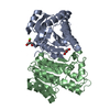
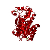
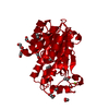
| Title | X-ray structure of Lactobacillus brevis alcohol dehydrogenase mutant Q126H | |||||||||
 Components Components | R-specific alcohol dehydrogenase | |||||||||
 Keywords Keywords | OXIDOREDUCTASE / Nucleotide Binding Oxidoreductase Activity | |||||||||
| Function / homology |  Function and homology information Function and homology informationbile acid metabolic process / oxidoreductase activity / nucleotide binding / metal ion binding Similarity search - Function | |||||||||
| Biological species |  Lactobacillus brevis (bacteria) Lactobacillus brevis (bacteria) | |||||||||
| Method |  X-RAY DIFFRACTION / SYNCHROTRON / MOLECULAR REPLACEMENT / Resolution: 1.22 Å X-RAY DIFFRACTION / SYNCHROTRON / MOLECULAR REPLACEMENT / Resolution: 1.22 Å | |||||||||
 Authors Authors | Hermann, J. / Bischoff, D. / Janowski, R. / Niessing, D. / Grob, P. / Hekmat, D. / Weuster-Botz, D. | |||||||||
| Funding support |  Germany, 1items Germany, 1items
| |||||||||
 Citation Citation | Journal: Crystals / Year: 2021 Title: Controlling Protein Crystallization by Free Energy Guided Design of Interactions at Crystal Contacts Authors: Hermann, J. / Bischoff, D. / Grob, P. / Janowski, R. / Hekmat, D. / Niessing, D. / Zacharias, M. / Weuster-Botz, D. | |||||||||
| History |
|
- Structure visualization
| Structure viewer | Molecule: MolmilJmol/JSmol |
|---|
- Downloads & links
-Download
| PDBx/mmCIF format | 6y10.cif.gz | 136.8 KB | Display | PDBx/mmCIF format |
|---|---|---|---|---|
| PDB format | pdb6y10.ent.gz | 104.7 KB | Display | PDB format |
| PDBx/mmJSON format | 6y10.json.gz | Tree view | PDBx/mmJSON format | |
| Others |  Other downloads Other downloads |
-Validation report
| Arichive directory | https://data.pdbj.org/pub/pdb/validation_reports/y1/6y10ftp://data.pdbj.org/pub/pdb/validation_reports/y1/6y10 | HTTPS FTP |
|---|
-Related structure data
| Related structure data | 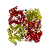 6y0zC 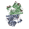 6y1cC  7a2bC 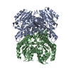 6h07S S: Starting model for refinement C: citing same article ( |
|---|---|
| Similar structure data |


-Links
 PDBj
PDBj


- Assembly
| Deposited unit | 
| ||||||||||||||||||||||||
|---|---|---|---|---|---|---|---|---|---|---|---|---|---|---|---|---|---|---|---|---|---|---|---|---|---|
| 1 | 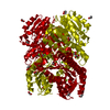
| ||||||||||||||||||||||||
| Unit cell |
| ||||||||||||||||||||||||
| Components on special symmetry positions |
|
-Components
| #1: Protein | Mass: 27884.389 Da / Num. of mol.: 1 Source method: isolated from a genetically manipulated source Source: (gene. exp.) Lactobacillus brevis (bacteria) / Gene: radh / Production host: | ||||||
|---|---|---|---|---|---|---|---|
| #2: Chemical | ChemComp-MG /   Mass: 24.305 Da / Num. of mol.: 4 / Source method: obtained synthetically / Formula: Mg Mass: 24.305 Da / Num. of mol.: 4 / Source method: obtained synthetically / Formula: Mg#3: Chemical | ChemComp-EDO /   Mass: 62.068 Da / Num. of mol.: 8 / Source method: obtained synthetically / Formula: C2H6O2 Mass: 62.068 Da / Num. of mol.: 8 / Source method: obtained synthetically / Formula: C2H6O2#4: Water | ChemComp-HOH / |  Mass: 18.015 Da / Num. of mol.: 407 / Source method: isolated from a natural source / Formula: H2O Mass: 18.015 Da / Num. of mol.: 407 / Source method: isolated from a natural source / Formula: H2OHas ligand of interest | N | |
-Experimental details
-Experiment
| Experiment | Method: X-RAY DIFFRACTION / Number of used crystals: 1 |
|---|
- Sample preparation
| Crystal | Density Matthews: 2.3 Å3/Da / Density % sol: 46.45 % |
|---|---|
| Crystal grow | Temperature: 293 K / Method: microbatch Details: Protein solution (10 g/L LbADH , 20 mM HEPES/NaOH pH 7.0, 1 mM MgCl2 and precipitation buffer (1 mM Tris/HCl pH 7.0, 50 mM MgCl2 and 100 g/L PEG 550 MME) |
-Data collection
| Diffraction | Mean temperature: 100 K / Serial crystal experiment: N |
|---|---|
| Diffraction source | Source: SYNCHROTRON / Site: SLS  / Beamline: X06DA / Wavelength: 0.966 Å / Beamline: X06DA / Wavelength: 0.966 Å |
| Detector | Type: DECTRIS PILATUS 2M-F / Detector: PIXEL / Date: Sep 12, 2019 |
| Radiation | Protocol: SINGLE WAVELENGTH / Monochromatic (M) / Laue (L): M / Scattering type: x-ray |
| Radiation wavelength | Wavelength: 0.966 Å / Relative weight: 1 |
| Reflection | Resolution: 1.22→46.01 Å / Num. obs: 75851 / % possible obs: 99.1 % / Redundancy: 6.96 % / CC1/2: 0.999 / Net I/σ(I): 13.49 |
| Reflection shell | Resolution: 1.22→1.25 Å / Num. unique obs: 5601 / CC1/2: 0.709 / % possible all: 99.4 |
- Processing
| Software |
| |||||||||||||||||||||||||||||||||||||||||||||||||||||||||||||||||
|---|---|---|---|---|---|---|---|---|---|---|---|---|---|---|---|---|---|---|---|---|---|---|---|---|---|---|---|---|---|---|---|---|---|---|---|---|---|---|---|---|---|---|---|---|---|---|---|---|---|---|---|---|---|---|---|---|---|---|---|---|---|---|---|---|---|---|
| Refinement | Method to determine structure: MOLECULAR REPLACEMENT Starting model: 6H07 Resolution: 1.22→46.01 Å / Cor.coef. Fo:Fc: 0.979 / Cor.coef. Fo:Fc free: 0.973 / SU B: 1.389 / SU ML: 0.026 / Cross valid method: THROUGHOUT / σ(F): 0 / ESU R: 0.039 / ESU R Free: 0.038 / Stereochemistry target values: MAXIMUM LIKELIHOOD Details: HYDROGENS HAVE BEEN ADDED IN THE RIDING POSITIONS U VALUES : REFINED INDIVIDUALLY
| |||||||||||||||||||||||||||||||||||||||||||||||||||||||||||||||||
| Solvent computation | Ion probe radii: 0.8 Å / Shrinkage radii: 0.8 Å / VDW probe radii: 1.2 Å / Solvent model: MASK | |||||||||||||||||||||||||||||||||||||||||||||||||||||||||||||||||
| Displacement parameters | Biso max: 70.59 Å2 / Biso mean: 14.309 Å2 / Biso min: 6.73 Å2
| |||||||||||||||||||||||||||||||||||||||||||||||||||||||||||||||||
| Refinement step | Cycle: final / Resolution: 1.22→46.01 Å
| |||||||||||||||||||||||||||||||||||||||||||||||||||||||||||||||||
| Refine LS restraints |
| |||||||||||||||||||||||||||||||||||||||||||||||||||||||||||||||||
| LS refinement shell | Resolution: 1.22→1.252 Å / Rfactor Rfree error: 0 / Total num. of bins used: 20
|
