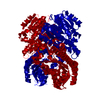
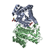
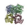
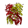
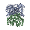
Entry Database : PDB / ID : 6h07Title X-ray structure of Lactobacillus brevis alcohol dehydrogenase R-specific alcohol dehydrogenase Keywords / / / Function / homology Function Domain/homology Component
/ / / / / / / / / Biological species Lactobacillus brevis (bacteria)Method / / / Resolution : 1.482 Å Authors Hermann, J. / Nowotny, P. / Biggel, P. / Schneider, S. / Hekmat, D. / Weuster-Botz, D. Funding support Organization Grant number Country German Research Foundation SPP 1934
Journal : Acta Crystallogr F Struct Biol Commun / Year : 2018Title : Neutron and X-ray crystal structures of Lactobacillus brevis alcohol dehydrogenase reveal new insights into hydrogen-bonding pathways.Authors : Hermann, J. / Nowotny, P. / Schrader, T.E. / Biggel, P. / Hekmat, D. / Weuster-Botz, D. History Deposition Jul 6, 2018 Deposition site / Processing site Revision 1.0 Dec 12, 2018 Provider / Type Revision 1.1 Nov 6, 2019 Group / Database references / Category / Item Revision 1.2 Jan 17, 2024 Group Data collection / Database references ... Data collection / Database references / Derived calculations / Refinement description Category chem_comp_atom / chem_comp_bond ... chem_comp_atom / chem_comp_bond / database_2 / pdbx_initial_refinement_model / pdbx_struct_conn_angle / refine_hist / struct_conn Item _database_2.pdbx_DOI / _database_2.pdbx_database_accession ... _database_2.pdbx_DOI / _database_2.pdbx_database_accession / _pdbx_struct_conn_angle.ptnr1_auth_asym_id / _pdbx_struct_conn_angle.ptnr1_auth_seq_id / _pdbx_struct_conn_angle.ptnr1_label_asym_id / _pdbx_struct_conn_angle.ptnr1_symmetry / _pdbx_struct_conn_angle.ptnr2_symmetry / _pdbx_struct_conn_angle.ptnr3_auth_asym_id / _pdbx_struct_conn_angle.ptnr3_auth_seq_id / _pdbx_struct_conn_angle.ptnr3_label_asym_id / _pdbx_struct_conn_angle.ptnr3_symmetry / _pdbx_struct_conn_angle.value / _refine_hist.d_res_high / _struct_conn.pdbx_dist_value / _struct_conn.pdbx_ptnr1_label_alt_id / _struct_conn.pdbx_ptnr2_label_alt_id / _struct_conn.ptnr1_auth_asym_id / _struct_conn.ptnr1_auth_comp_id / _struct_conn.ptnr1_auth_seq_id / _struct_conn.ptnr1_label_asym_id / _struct_conn.ptnr1_label_atom_id / _struct_conn.ptnr1_label_comp_id / _struct_conn.ptnr1_symmetry / _struct_conn.ptnr2_auth_asym_id / _struct_conn.ptnr2_auth_comp_id / _struct_conn.ptnr2_auth_seq_id / _struct_conn.ptnr2_label_asym_id / _struct_conn.ptnr2_label_atom_id / _struct_conn.ptnr2_label_comp_id / _struct_conn.ptnr2_symmetry
Show all Show less
 Movie
Movie Controller


 Open data
 Basic information
 Components
 Keywords
 Function and homology information
 Lactobacillus brevis (bacteria)
 X-RAY DIFFRACTION /
  Authors
 Germany, 1items
  Citation
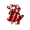
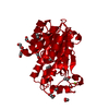
 Structure visualization
 Downloads & links
 Other downloads
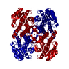


 PDBj
PDBj


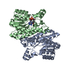
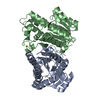
 Assembly

 Mass: 24.305 Da / Num. of mol.: 1 / Source method: obtained synthetically / Formula: Mg
 Mass: 54.938 Da / Num. of mol.: 1 / Source method: isolated from a natural source / Formula: Mn
 Mass: 18.015 Da / Num. of mol.: 391 / Source method: isolated from a natural source / Formula: H2O
 Sample preparation
 / Beamline: MASSIF-3 / Wavelength: 0.968 Å
 Processing