[English] 日本語
 Yorodumi
- PDB-6xze: crystal structure of human carbonic anhydrase I in complex with 4... -
+ Open data
- Basic information
| Entry | Database: PDB / ID: 6xze | ||||||
|---|---|---|---|---|---|---|---|
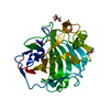
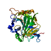
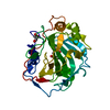
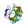
| Title | crystal structure of human carbonic anhydrase I in complex with 4-(3-(2-((2-fluorobenzyl)amino)ethyl)ureido) benzenesulfonamide | ||||||
 Components Components | Carbonic anhydrase 1 | ||||||
 Keywords Keywords | LYASE / Inhibitor / carbon dioxide / carbonic anhydrase | ||||||
| Function / homology |  Function and homology information Function and homology informationhydro-lyase activity / cyanamide hydratase / cyanamide hydratase activity / arylesterase activity / Gene and protein expression by JAK-STAT signaling after Interleukin-12 stimulation / Reversible hydration of carbon dioxide / carbonic anhydrase / carbonate dehydratase activity / Erythrocytes take up oxygen and release carbon dioxide / Erythrocytes take up carbon dioxide and release oxygen ...hydro-lyase activity / cyanamide hydratase / cyanamide hydratase activity / arylesterase activity / Gene and protein expression by JAK-STAT signaling after Interleukin-12 stimulation / Reversible hydration of carbon dioxide / carbonic anhydrase / carbonate dehydratase activity / Erythrocytes take up oxygen and release carbon dioxide / Erythrocytes take up carbon dioxide and release oxygen / extracellular exosome / zinc ion binding / cytoplasm / cytosol Similarity search - Function | ||||||
| Biological species |  Homo sapiens (human) Homo sapiens (human) | ||||||
| Method |  X-RAY DIFFRACTION / X-RAY DIFFRACTION /  SYNCHROTRON / SYNCHROTRON /  MOLECULAR REPLACEMENT / Resolution: 1.54 Å MOLECULAR REPLACEMENT / Resolution: 1.54 Å | ||||||
 Authors Authors | Zanotti, G. / Majid, A. / Bozdag, M. / Angeli, A. / Carta, F. / Berto, P. / Supuran, C. | ||||||
 Citation Citation |  Journal: Int J Mol Sci / Year: 2020 Journal: Int J Mol Sci / Year: 2020Title: Benzylaminoethyureido-Tailed Benzenesulfonamides: Design, Synthesis, Kinetic and X-ray Investigations on Human Carbonic Anhydrases. Authors: Ali, M. / Bozdag, M. / Farooq, U. / Angeli, A. / Carta, F. / Berto, P. / Zanotti, G. / Supuran, C.T. | ||||||
| History |
|
- Structure visualization
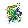
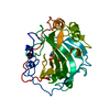
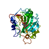
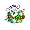
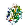
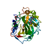
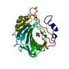
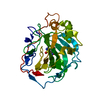
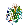
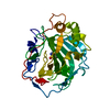
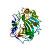
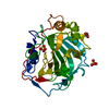
| Structure viewer | Molecule:  Molmil Molmil Jmol/JSmol Jmol/JSmol |
|---|
- Downloads & links
- Download
| PDBx/mmCIF format |  6xze.cif.gz 6xze.cif.gz | 132.9 KB | Display |  PDBx/mmCIF format PDBx/mmCIF format |
|---|---|---|---|---|
| PDB format |  pdb6xze.ent.gz pdb6xze.ent.gz | 100.3 KB | Display |  PDB format PDB format |
| PDBx/mmJSON format |  6xze.json.gz 6xze.json.gz | Tree view |  PDBx/mmJSON format PDBx/mmJSON format | |
| Others |  Other downloads Other downloads |
-Validation report
| Arichive directory |  https://data.pdbj.org/pub/pdb/validation_reports/xz/6xze https://data.pdbj.org/pub/pdb/validation_reports/xz/6xze ftp://data.pdbj.org/pub/pdb/validation_reports/xz/6xze ftp://data.pdbj.org/pub/pdb/validation_reports/xz/6xze | HTTPS FTP |
|---|
-Related structure data
| Related structure data | 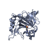 6xzoC 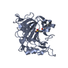 6xzsC 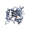 6xzxC 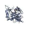 6xzyC 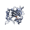 6y00C  6g3vS S: Starting model for refinement C: citing same article ( |
|---|---|
| Similar structure data |
- Links
Links
- Assembly
| Deposited unit | 
| ||||||||||||
|---|---|---|---|---|---|---|---|---|---|---|---|---|---|
| 1 | 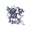
| ||||||||||||
| 2 | 
| ||||||||||||
| Unit cell |
|
- Components
| #1: Protein | Mass: 28906.186 Da / Num. of mol.: 2 / Source method: isolated from a natural source / Source: (natural)  Homo sapiens (human) / Tissue: erythrocytes / References: UniProt: P00915, carbonic anhydrase Homo sapiens (human) / Tissue: erythrocytes / References: UniProt: P00915, carbonic anhydrase#2: Chemical | #3: Chemical | #4: Water | ChemComp-HOH / | Has ligand of interest | Y | |
|---|
-Experimental details
-Experiment
| Experiment | Method:  X-RAY DIFFRACTION / Number of used crystals: 1 X-RAY DIFFRACTION / Number of used crystals: 1 |
|---|
- Sample preparation
| Crystal | Density Matthews: 2.36 Å3/Da / Density % sol: 47.8 % |
|---|---|
| Crystal grow | Temperature: 293 K / Method: vapor diffusion, sitting drop / pH: 9 Details: 200 mM Na-acetate, 30% PEG 4000, 100 mM Tris-HCl Inhibitor: 10 mM inhibitor solution (150 mM NaCl, 10% DMSO, 50 mM Tris pH 7) |
-Data collection
| Diffraction | Mean temperature: 100 K / Serial crystal experiment: N |
|---|---|
| Diffraction source | Source:  SYNCHROTRON / Site: SYNCHROTRON / Site:  Diamond Diamond  / Beamline: I03 / Wavelength: 0.976 Å / Beamline: I03 / Wavelength: 0.976 Å |
| Detector | Type: DECTRIS EIGER2 XE 16M / Detector: PIXEL / Date: Jul 4, 2019 |
| Radiation | Protocol: SINGLE WAVELENGTH / Monochromatic (M) / Laue (L): M / Scattering type: x-ray |
| Radiation wavelength | Wavelength: 0.976 Å / Relative weight: 1 |
| Reflection | Resolution: 1.54→62.6 Å / Num. obs: 81860 / % possible obs: 100 % / Observed criterion σ(F): 0 / Observed criterion σ(I): 0 / Redundancy: 13.2 % / Rmerge(I) obs: 0.103 / Rpim(I) all: 0.029 / Net I/σ(I): 11.4 |
| Reflection shell | Resolution: 1.54→1.58 Å / Redundancy: 13.4 % / Rmerge(I) obs: 1.8 / Mean I/σ(I) obs: 1.2 / Num. unique obs: 5955 / Rpim(I) all: 0.557 / % possible all: 100 |
- Processing
| Software |
| ||||||||||||||||||||||||
|---|---|---|---|---|---|---|---|---|---|---|---|---|---|---|---|---|---|---|---|---|---|---|---|---|---|
| Refinement | Method to determine structure:  MOLECULAR REPLACEMENT MOLECULAR REPLACEMENTStarting model: 6g3v Resolution: 1.54→55.667 Å / SU ML: 0.19 / Cross valid method: FREE R-VALUE / Phase error: 26.22
| ||||||||||||||||||||||||
| Solvent computation | Shrinkage radii: 0.9 Å / VDW probe radii: 1.11 Å | ||||||||||||||||||||||||
| Displacement parameters | Biso mean: 28.6 Å2 | ||||||||||||||||||||||||
| Refinement step | Cycle: LAST / Resolution: 1.54→55.667 Å
| ||||||||||||||||||||||||
| Refine LS restraints |
|
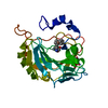
 Movie
Movie Controller



 PDBj
PDBj


