| 登録情報 | データベース: PDB / ID: 6xx9
|
|---|
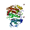
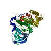
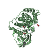
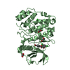
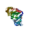
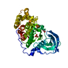
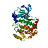
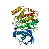
| タイトル | Arabidopsis thaliana Casein Kinase 2 (CK2) alpha-1 crystal form III |
|---|
 要素 要素 | Casein kinase II subunit alpha-1 |
|---|
 キーワード キーワード | CELL CYCLE / Kinase / CK2 / Casein Kinase |
|---|
| 機能・相同性 |  機能・相同性情報 機能・相同性情報
response to UV-C / protein kinase CK2 complex / response to gamma radiation / circadian rhythm / regulation of circadian rhythm / kinase activity / chromatin organization / non-specific serine/threonine protein kinase / regulation of cell cycle / protein serine kinase activity ...response to UV-C / protein kinase CK2 complex / response to gamma radiation / circadian rhythm / regulation of circadian rhythm / kinase activity / chromatin organization / non-specific serine/threonine protein kinase / regulation of cell cycle / protein serine kinase activity / DNA repair / protein serine/threonine kinase activity / DNA damage response / nucleolus / ATP binding / nucleus / cytosol類似検索 - 分子機能 Casein Kinase 2, subunit alpha / Phosphorylase Kinase; domain 1 / Phosphorylase Kinase; domain 1 / Transferase(Phosphotransferase) domain 1 / Transferase(Phosphotransferase); domain 1 / Serine/threonine-protein kinase, active site / Serine/Threonine protein kinases active-site signature. / Protein kinase domain / Serine/Threonine protein kinases, catalytic domain / Protein kinase, ATP binding site ...Casein Kinase 2, subunit alpha / Phosphorylase Kinase; domain 1 / Phosphorylase Kinase; domain 1 / Transferase(Phosphotransferase) domain 1 / Transferase(Phosphotransferase); domain 1 / Serine/threonine-protein kinase, active site / Serine/Threonine protein kinases active-site signature. / Protein kinase domain / Serine/Threonine protein kinases, catalytic domain / Protein kinase, ATP binding site / Protein kinases ATP-binding region signature. / Protein kinase domain profile. / Protein kinase domain / Protein kinase-like domain superfamily / 2-Layer Sandwich / Orthogonal Bundle / Mainly Alpha / Alpha Beta類似検索 - ドメイン・相同性 |
|---|
| 生物種 |   Arabidopsis thaliana (シロイヌナズナ) Arabidopsis thaliana (シロイヌナズナ) |
|---|
| 手法 |  X線回折 / シンクロトロン / 分子置換 / 解像度: 1.84 Å X線回折 / シンクロトロン / 分子置換 / 解像度: 1.84 Å |
|---|
 データ登録者 データ登録者 | Demulder, M. / De Veylder, L. / Loris, R. |
|---|
| 資金援助 |  ベルギー, 2件 ベルギー, 2件 | 組織 | 認可番号 | 国 |
|---|
| Research Foundation - Flanders (FWO) | G023616N | ベルギー | | Research Foundation - Flanders (FWO) | G011420N | ベルギー |
|
|---|
 引用 引用 | ジャーナル: Acta Crystallogr.,Sect.F / 年: 2020
タイトル: Crystal structure of Arabidopsis thaliana casein kinase 2 alpha 1.
著者: Demulder, M. / De Veylder, L. / Loris, R. |
|---|
| 履歴 | | 登録 | 2020年1月27日 | 登録サイト: PDBE / 処理サイト: PDBE |
|---|
| 改定 1.0 | 2020年4月15日 | Provider: repository / タイプ: Initial release |
|---|
| 改定 1.1 | 2024年1月24日 | Group: Data collection / Database references / Refinement description
カテゴリ: chem_comp_atom / chem_comp_bond ...chem_comp_atom / chem_comp_bond / database_2 / pdbx_initial_refinement_model
Item: _database_2.pdbx_DOI / _database_2.pdbx_database_accession |
|---|
|
|---|
 ムービー
ムービー コントローラー


 データを開く
 基本情報
基本情報 構造の表示
構造の表示 ダウンロードとリンク
 その他のダウンロード


 PDBj
PDBj 集合体
集合体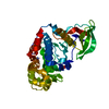


 分子量: 122.143 Da / 分子数: 1 / 由来タイプ: 合成 / 式: C4H12NO3 / コメント: pH緩衝剤*YM
 分子量: 96.063 Da / 分子数: 2 / 由来タイプ: 合成 / 式: SO4
 分子量: 18.015 Da / 分子数: 180 / 由来タイプ: 天然 / 式: H2O
 試料調製
試料調製 / ビームライン: PROXIMA 2 / 波長: 0.9801 Å
 解析
解析