| Entry | Database: PDB / ID: 6xuv
|
|---|
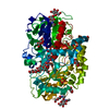
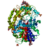
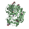
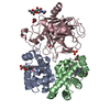
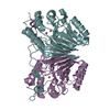
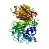
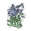
| Title | Crystallographic structure of oligosaccharide dehydrogenase from Pycnoporus cinnabarinus, laminaribiose-bound form |
|---|
 Components Components | Oligosaccharide dehydrogenase |
|---|
 Keywords Keywords | OXIDOREDUCTASE / Glucose dehydrogenase / Pycnoporus cinnabarinus / Laminaribiose / oligosaccharide dehydrogenase |
|---|
| Function / homology |  Function and homology information Function and homology information
Glucose Oxidase, domain 2 / GMC oxidoreductases signature 1. / GMC oxidoreductases signature 2. / Glucose-methanol-choline oxidoreductase / Glucose-methanol-choline oxidoreductase, N-terminal / GMC oxidoreductase / Glucose-methanol-choline oxidoreductase, C-terminal / GMC oxidoreductase / FAD/NAD(P)-binding domain superfamilySimilarity search - Domain/homology |
|---|
| Biological species |  Trametes cinnabarina (fungus) Trametes cinnabarina (fungus) |
|---|
| Method |  X-RAY DIFFRACTION / SYNCHROTRON / FOURIER SYNTHESIS / Resolution: 1.75 Å X-RAY DIFFRACTION / SYNCHROTRON / FOURIER SYNTHESIS / Resolution: 1.75 Å |
|---|
 Authors Authors | Cerutti, G. / Savino, C. / Montemiglio, L.C. / Vallone, B. / Sciara, G. |
|---|
| Funding support |  France, 2items France, 2items | Organization | Grant number | Country |
|---|
| French National Research Agency | ANR 19-CE43-0007 | France | | French National Institute of Agricultural Research (INRAE) | ANS 2018-2019 | France |
|
|---|
 Citation Citation | Journal: Biotechnol Biofuels / Year: 2021
Title: Crystal structure and functional characterization of an oligosaccharide dehydrogenase from Pycnoporus cinnabarinus provides insights into fungal breakdown of lignocellulose.
Authors: Cerutti, G. / Gugole, E. / Montemiglio, L.C. / Turbe-Doan, A. / Chena, D. / Navarro, D. / Lomascolo, A. / Piumi, F. / Exertier, C. / Freda, I. / Vallone, B. / Record, E. / Savino, C. / Sciara, G. |
|---|
| History | | Deposition | Jan 21, 2020 | Deposition site: PDBE / Processing site: PDBE |
|---|
| Revision 1.0 | Feb 3, 2021 | Provider: repository / Type: Initial release |
|---|
| Revision 1.1 | Aug 11, 2021 | Group: Database references / Category: citation / citation_author / database_2
Item: _citation.country / _citation.journal_abbrev ..._citation.country / _citation.journal_abbrev / _citation.journal_id_CSD / _citation.journal_id_ISSN / _citation.journal_volume / _citation.page_first / _citation.page_last / _citation.pdbx_database_id_DOI / _citation.pdbx_database_id_PubMed / _citation.title / _citation.year / _database_2.pdbx_DOI / _database_2.pdbx_database_accession |
|---|
| Revision 1.2 | Nov 20, 2024 | Group: Data collection / Structure summary
Category: chem_comp_atom / chem_comp_bond ...chem_comp_atom / chem_comp_bond / pdbx_entity_branch_descriptor / pdbx_entry_details / pdbx_modification_feature
Item: _pdbx_entry_details.has_protein_modification |
|---|
|
|---|
 Movie
Movie Controller

 Yorodumi
 Open data
 Basic information
 Structure visualization
 Downloads & links
 Other downloads

 PDBj
PDBj


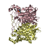
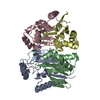
 Assembly
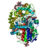


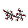

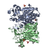
 Type: D-saccharide, alpha linking / Mass: 180.156 Da / Num. of mol.: 1
 Type: D-saccharide, beta linking / Mass: 221.208 Da / Num. of mol.: 1


 Mass: 785.550 Da / Num. of mol.: 1 / Source method: obtained synthetically / Formula: C27H33N9O15P2 / Feature type: SUBJECT OF INVESTIGATION / Comment: FAD*YM
 Mass: 96.063 Da / Num. of mol.: 3 / Source method: obtained synthetically / Formula: SO4
 Sample preparation
 / Beamline: I24 / Wavelength: 0.9685 Å
 Processing