 Movie
Movie Controller

[English] 日本語
 Yorodumi
- PDB-6xqf: Crystal structure of SCLam E144S mutant, a non-specific endo-beta... -
+ Open data

- Basic information
| Entry | Database: PDB / ID: 6xqf | ||||||||||||
|---|---|---|---|---|---|---|---|---|---|---|---|---|---|
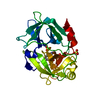
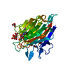
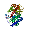
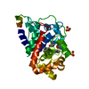
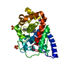
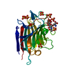
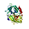
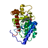
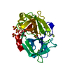
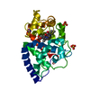
| Title | Crystal structure of SCLam E144S mutant, a non-specific endo-beta-1,3(4)-glucanase from family GH16, co-crystallized with 1,3-beta-D-cellotriosyl-glucose, presenting a 1,3-beta-D-cellobiosyl-glucose at active site | ||||||||||||
 Components Components | GH16 family protein | ||||||||||||
 Keywords Keywords | HYDROLASE / glycoside hydrolase / transglycosylation / endo-1 / 3(4)-beta-glucanases / metagenome | ||||||||||||
| Function / homology |  Function and homology information Function and homology informationglucan endo-1,3-beta-D-glucosidase / glucan endo-1,3-beta-D-glucosidase activity / carbohydrate metabolic process / metal ion binding Similarity search - Function | ||||||||||||
| Biological species |  uncultured bacterium (environmental samples) uncultured bacterium (environmental samples) | ||||||||||||
| Method |  X-RAY DIFFRACTION / SYNCHROTRON / MOLECULAR REPLACEMENT / molecular replacement / Resolution: 1.58 Å X-RAY DIFFRACTION / SYNCHROTRON / MOLECULAR REPLACEMENT / molecular replacement / Resolution: 1.58 Å | ||||||||||||
 Authors Authors | Liberato, M.V. / Squina, F. | ||||||||||||
| Funding support |  Brazil, 3items Brazil, 3items
| ||||||||||||
 Citation Citation | Journal: J.Biol.Chem. / Year: 2021 Title: Insights into the dual cleavage activity of the GH16 laminarinase enzyme class on beta-1,3 and beta-1,4 glycosidic bonds. Authors: Liberato, M.V. / Teixeira Prates, E. / Goncalves, T.A. / Bernardes, A. / Vilela, N. / Fattori, J. / Ematsu, G.C. / Chinaglia, M. / Machi Gomes, E.R. / Migliorini Figueira, A.C. / Damasio, A. ...Authors: Liberato, M.V. / Teixeira Prates, E. / Goncalves, T.A. / Bernardes, A. / Vilela, N. / Fattori, J. / Ematsu, G.C. / Chinaglia, M. / Machi Gomes, E.R. / Migliorini Figueira, A.C. / Damasio, A. / Polikarpov, I. / Skaf, M.S. / Squina, F.M. | ||||||||||||
| History |
|
- Structure visualization
| Structure viewer | Molecule: MolmilJmol/JSmol |
|---|
- Downloads & links
-Download
| PDBx/mmCIF format | 6xqf.cif.gz | 139.5 KB | Display | PDBx/mmCIF format |
|---|---|---|---|---|
| PDB format | pdb6xqf.ent.gz | 105.3 KB | Display | PDB format |
| PDBx/mmJSON format | 6xqf.json.gz | Tree view | PDBx/mmJSON format | |
| Others |  Other downloads Other downloads |
-Validation report
| Arichive directory | https://data.pdbj.org/pub/pdb/validation_reports/xq/6xqfftp://data.pdbj.org/pub/pdb/validation_reports/xq/6xqf | HTTPS FTP |
|---|
-Related structure data
| Related structure data | 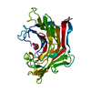 6xofSC 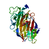 6xqgC 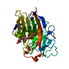 6xqhC  6xqlC  6xqmC S: Starting model for refinement C: citing same article ( |
|---|---|
| Similar structure data |
-Links
 PDBj
PDBj
- Assembly
| Deposited unit | 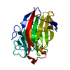
| ||||||||
|---|---|---|---|---|---|---|---|---|---|
| 1 |
| ||||||||
| Unit cell |
|
-Components
| #1: Protein | Mass: 30288.746 Da / Num. of mol.: 1 / Mutation: E144S Source method: isolated from a genetically manipulated source Source: (gene. exp.) uncultured bacterium (environmental samples)Gene: sclam / Production host: References: UniProt: A0A0B5H9B3, glucan endo-1,3-beta-D-glucosidase |
|---|---|
| #2: Polysaccharide | beta-D-glucopyranose-(1-4)-beta-D-glucopyranose-(1-3)-alpha-D-glucopyranose Source method: isolated from a genetically manipulated source |
| #3: Chemical | ChemComp-CA /   Mass: 40.078 Da / Num. of mol.: 1 / Source method: obtained synthetically / Formula: Ca / Feature type: SUBJECT OF INVESTIGATION Mass: 40.078 Da / Num. of mol.: 1 / Source method: obtained synthetically / Formula: Ca / Feature type: SUBJECT OF INVESTIGATION |
| #4: Water | ChemComp-HOH /  Mass: 18.015 Da / Num. of mol.: 455 / Source method: isolated from a natural source / Formula: H2O Mass: 18.015 Da / Num. of mol.: 455 / Source method: isolated from a natural source / Formula: H2O |
| Has ligand of interest | Y |
| Has protein modification | Y |
-Experimental details
-Experiment
| Experiment | Method: X-RAY DIFFRACTION / Number of used crystals: 1 |
|---|
- Sample preparation
| Crystal | Density Matthews: 1.85 Å3/Da / Density % sol: 33.45 % |
|---|---|
| Crystal grow | Temperature: 291 K / Method: vapor diffusion, hanging drop / pH: 8.5 Details: 30 % PEG4000, 0.2 M magnesium chloride, and 0.1 M Tris |
-Data collection
| Diffraction | Mean temperature: 100 K / Serial crystal experiment: N | ||||||||||||||||||||||||||||||
|---|---|---|---|---|---|---|---|---|---|---|---|---|---|---|---|---|---|---|---|---|---|---|---|---|---|---|---|---|---|---|---|
| Diffraction source | Source: SYNCHROTRON / Site: LNLS / Beamline: W01B-MX2 / Wavelength: 1.4583 Å | ||||||||||||||||||||||||||||||
| Detector | Type: DECTRIS PILATUS 2M / Detector: PIXEL / Date: Mar 25, 2015 | ||||||||||||||||||||||||||||||
| Radiation | Protocol: SINGLE WAVELENGTH / Monochromatic (M) / Laue (L): M / Scattering type: x-ray | ||||||||||||||||||||||||||||||
| Radiation wavelength | Wavelength: 1.4583 Å / Relative weight: 1 | ||||||||||||||||||||||||||||||
| Reflection | Resolution: 1.58→45.72 Å / Num. obs: 30357 / % possible obs: 97.2 % / Redundancy: 5 % / CC1/2: 0.989 / Rmerge(I) obs: 0.084 / Rpim(I) all: 0.042 / Rrim(I) all: 0.094 / Net I/σ(I): 13.9 / Num. measured all: 151100 / Scaling rejects: 832 | ||||||||||||||||||||||||||||||
| Reflection shell | Diffraction-ID: 1
|
-Phasing
| Phasing | Method: molecular replacement | |||||||||
|---|---|---|---|---|---|---|---|---|---|---|
| Phasing MR | Model details: Phaser MODE: MR_AUTO
|
- Processing
| Software |
| ||||||||||||||||||||||||||||||||||||||||||||||||||||||||||||
|---|---|---|---|---|---|---|---|---|---|---|---|---|---|---|---|---|---|---|---|---|---|---|---|---|---|---|---|---|---|---|---|---|---|---|---|---|---|---|---|---|---|---|---|---|---|---|---|---|---|---|---|---|---|---|---|---|---|---|---|---|---|
| Refinement | Method to determine structure: MOLECULAR REPLACEMENT Starting model: 6XOF Resolution: 1.58→45.72 Å / Cor.coef. Fo:Fc: 0.952 / Cor.coef. Fo:Fc free: 0.928 / WRfactor Rfree: 0.1899 / WRfactor Rwork: 0.1619 / FOM work R set: 0.8378 / SU B: 4.956 / SU ML: 0.076 / SU R Cruickshank DPI: 0.1034 / SU Rfree: 0.1012 / Cross valid method: THROUGHOUT / σ(F): 0 / ESU R: 0.103 / ESU R Free: 0.101 / Stereochemistry target values: MAXIMUM LIKELIHOOD Details: HYDROGENS HAVE BEEN ADDED IN THE RIDING POSITIONS U VALUES : WITH TLS ADDED
| ||||||||||||||||||||||||||||||||||||||||||||||||||||||||||||
| Solvent computation | Ion probe radii: 0.8 Å / Shrinkage radii: 0.8 Å / VDW probe radii: 1.2 Å / Solvent model: MASK | ||||||||||||||||||||||||||||||||||||||||||||||||||||||||||||
| Displacement parameters | Biso max: 48.99 Å2 / Biso mean: 10.568 Å2 / Biso min: 2.78 Å2
| ||||||||||||||||||||||||||||||||||||||||||||||||||||||||||||
| Refinement step | Cycle: final / Resolution: 1.58→45.72 Å
| ||||||||||||||||||||||||||||||||||||||||||||||||||||||||||||
| Refine LS restraints |
| ||||||||||||||||||||||||||||||||||||||||||||||||||||||||||||
| LS refinement shell | Resolution: 1.58→1.621 Å / Rfactor Rfree error: 0 / Total num. of bins used: 20
| ||||||||||||||||||||||||||||||||||||||||||||||||||||||||||||
| Refinement TLS params. | Method: refined / Origin x: -8.46 Å / Origin y: -4.624 Å / Origin z: 12.934 Å
| ||||||||||||||||||||||||||||||||||||||||||||||||||||||||||||
| Refinement TLS group |
|
