 Movie
Movie Controller


+ Open data

- Basic information
| Entry | Database: PDB / ID: 6xkd | ||||||
|---|---|---|---|---|---|---|---|
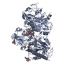
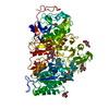
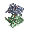
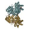
| Title | Structure of ligand-bound mouse cGAMP hydrolase ENPP1 | ||||||
 Components Components | Ectonucleotide pyrophosphatase/phosphodiesterase family member 1 | ||||||
 Keywords Keywords | HYDROLASE/HYDROLASE INHIBITOR / hydrolase / inhibitor / HYDROLASE-HYDROLASE INHIBITOR complex | ||||||
| Function / homology |  Function and homology information Function and homology informationphosphodiesterase I / nucleotide diphosphatase / hydrolase activity, acting on ester bonds / scavenger receptor activity / polysaccharide binding / nucleic acid binding / immune response / extracellular region / metal ion binding / membrane Similarity search - Function | ||||||
| Biological species |  | ||||||
| Method |  X-RAY DIFFRACTION / SYNCHROTRON / MOLECULAR REPLACEMENT / molecular replacement / Resolution: 3.2 Å X-RAY DIFFRACTION / SYNCHROTRON / MOLECULAR REPLACEMENT / molecular replacement / Resolution: 3.2 Å | ||||||
 Authors Authors | Fernandez, D. / Li, L. | ||||||
 Citation Citation | Journal: Cell Chem Biol / Year: 2020 Title: Structure-Aided Development of Small-Molecule Inhibitors of ENPP1, the Extracellular Phosphodiesterase of the Immunotransmitter cGAMP. Authors: Carozza, J.A. / Brown, J.A. / Bohnert, V. / Fernandez, D. / AlSaif, Y. / Mardjuki, R.E. / Smith, M. / Li, L. | ||||||
| History |
|
- Structure visualization
| Structure viewer | Molecule: MolmilJmol/JSmol |
|---|
- Downloads & links
-Download
| PDBx/mmCIF format | 6xkd.cif.gz | 291.6 KB | Display | PDBx/mmCIF format |
|---|---|---|---|---|
| PDB format | pdb6xkd.ent.gz | 227.3 KB | Display | PDB format |
| PDBx/mmJSON format | 6xkd.json.gz | Tree view | PDBx/mmJSON format | |
| Others |  Other downloads Other downloads |
-Validation report
| Summary document | 6xkd_validation.pdf.gz | 1.2 MB | Display | wwPDB validaton report |
|---|---|---|---|---|
| Full document | 6xkd_full_validation.pdf.gz | 1.2 MB | Display | |
| Data in XML | 6xkd_validation.xml.gz | 48.5 KB | Display | |
| Data in CIF | 6xkd_validation.cif.gz | 66.6 KB | Display | |
| Arichive directory | https://data.pdbj.org/pub/pdb/validation_reports/xk/6xkdftp://data.pdbj.org/pub/pdb/validation_reports/xk/6xkd | HTTPS FTP |
-Related structure data
| Related structure data |  4gtwS S: Starting model for refinement |
|---|---|
| Similar structure data |


-Links
 PDBj
PDBj
- Assembly
| Deposited unit | 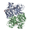
| ||||||||
|---|---|---|---|---|---|---|---|---|---|
| 1 | 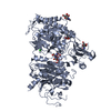
| ||||||||
| 2 | 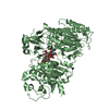
| ||||||||
| Unit cell |
|
-Components
-Protein , 1 types, 2 molecules AB
| #1: Protein | Mass: 94560.203 Da / Num. of mol.: 2 Source method: isolated from a genetically manipulated source Source: (gene. exp.)  Homo sapiens (human) / References: UniProt: G3X9S2 Homo sapiens (human) / References: UniProt: G3X9S2 |
|---|
-Sugars , 2 types, 4 molecules 
| #2: Polysaccharide | Source method: isolated from a genetically manipulated source #3: Sugar |  Type: D-saccharide, beta linking / Mass: 221.208 Da / Num. of mol.: 2 Type: D-saccharide, beta linking / Mass: 221.208 Da / Num. of mol.: 2Source method: isolated from a genetically manipulated source Formula: C8H15NO6 |
|---|
-Non-polymers , 5 types, 40 molecules 


| #4: Chemical | ChemComp-ZN /  Mass: 65.409 Da / Num. of mol.: 4 Mass: 65.409 Da / Num. of mol.: 4Source method: isolated from a genetically manipulated source Formula: Zn #5: Chemical |  Mass: 40.078 Da / Num. of mol.: 2 / Source method: obtained synthetically / Formula: Ca Mass: 40.078 Da / Num. of mol.: 2 / Source method: obtained synthetically / Formula: Ca#6: Chemical | ChemComp-CL / |  Mass: 35.453 Da / Num. of mol.: 1 / Source method: obtained synthetically / Formula: Cl Mass: 35.453 Da / Num. of mol.: 1 / Source method: obtained synthetically / Formula: Cl#7: Chemical | ChemComp-IJE / { |  Mass: 381.363 Da / Num. of mol.: 1 / Source method: obtained synthetically / Formula: C17H24N3O5P / Feature type: SUBJECT OF INVESTIGATION Mass: 381.363 Da / Num. of mol.: 1 / Source method: obtained synthetically / Formula: C17H24N3O5P / Feature type: SUBJECT OF INVESTIGATION#8: Water | ChemComp-HOH / | Mass: 18.015 Da / Num. of mol.: 32 / Source method: isolated from a natural source / Formula: H2O |
|---|
-Details
| Has ligand of interest | Y |
|---|---|
| Has protein modification | Y |
| Nonpolymer details | The authors state that the zinc-binding group and hydrophobic core of the inhibitor have weak ...The authors state that the zinc-binding group and hydrophobic core of the inhibitor have weak density in the binding pocket of ENPP1. The piperidinyl linker moiety shows very weak density apparently because of high flexibility of the hydrocarbon and it being exposed to the solvent. In polypeptide chain A the inhibitor was modeled with 0.5 occupancy. Trace of inhibitor in the electron density appears to be present in polypeptide B chain but the density is very weak and the inhibitor wasn't modeled there. |
-Experimental details
-Experiment
| Experiment | Method: X-RAY DIFFRACTION / Number of used crystals: 1 |
|---|
- Sample preparation
| Crystal | Density Matthews: 2.76 Å3/Da / Density % sol: 55.51 % / Description: Needle |
|---|---|
| Crystal grow | Temperature: 295 K / Method: vapor diffusion, hanging drop / pH: 4.5 Details: PEG 600, sodium acetate, magnesium acetate, polyvinylpyrrolidone |
-Data collection
| Diffraction | Mean temperature: 100 K / Serial crystal experiment: N | |||||||||||||||||||||||||||||||||||||||||||||||||||||||||||||||||||||||||||||||||||||||||||||||||||||||||||||||||||||||||
|---|---|---|---|---|---|---|---|---|---|---|---|---|---|---|---|---|---|---|---|---|---|---|---|---|---|---|---|---|---|---|---|---|---|---|---|---|---|---|---|---|---|---|---|---|---|---|---|---|---|---|---|---|---|---|---|---|---|---|---|---|---|---|---|---|---|---|---|---|---|---|---|---|---|---|---|---|---|---|---|---|---|---|---|---|---|---|---|---|---|---|---|---|---|---|---|---|---|---|---|---|---|---|---|---|---|---|---|---|---|---|---|---|---|---|---|---|---|---|---|---|---|---|
| Diffraction source | Source: SYNCHROTRON / Site: ALS  / Beamline: 5.0.1 / Wavelength: 0.97741 Å / Beamline: 5.0.1 / Wavelength: 0.97741 Å | |||||||||||||||||||||||||||||||||||||||||||||||||||||||||||||||||||||||||||||||||||||||||||||||||||||||||||||||||||||||||
| Detector | Type: DECTRIS PILATUS3 S 6M / Detector: PIXEL / Date: Sep 22, 2017 | |||||||||||||||||||||||||||||||||||||||||||||||||||||||||||||||||||||||||||||||||||||||||||||||||||||||||||||||||||||||||
| Radiation | Protocol: SINGLE WAVELENGTH / Monochromatic (M) / Laue (L): M / Scattering type: x-ray | |||||||||||||||||||||||||||||||||||||||||||||||||||||||||||||||||||||||||||||||||||||||||||||||||||||||||||||||||||||||||
| Radiation wavelength | Wavelength: 0.97741 Å / Relative weight: 1 | |||||||||||||||||||||||||||||||||||||||||||||||||||||||||||||||||||||||||||||||||||||||||||||||||||||||||||||||||||||||||
| Reflection | Resolution: 3.2→88.638 Å / Num. all: 30691 / Num. obs: 30691 / % possible obs: 91.8 % / Redundancy: 2.1 % / Rpim(I) all: 0.124 / Rrim(I) all: 0.204 / Rsym value: 0.16 / Net I/av σ(I): 3.8 / Net I/σ(I): 5.4 / Num. measured all: 65417 | |||||||||||||||||||||||||||||||||||||||||||||||||||||||||||||||||||||||||||||||||||||||||||||||||||||||||||||||||||||||||
| Reflection shell | Diffraction-ID: 1
|
-Phasing
| Phasing | Method: molecular replacement |
|---|
- Processing
| Software |
| |||||||||||||||||||||||||||||||||||||||||||||
|---|---|---|---|---|---|---|---|---|---|---|---|---|---|---|---|---|---|---|---|---|---|---|---|---|---|---|---|---|---|---|---|---|---|---|---|---|---|---|---|---|---|---|---|---|---|---|
| Refinement | Method to determine structure: MOLECULAR REPLACEMENT Starting model: 4gtw Resolution: 3.2→30.64 Å / Cor.coef. Fo:Fc: 0.923 / Cor.coef. Fo:Fc free: 0.848 / SU B: 28.054 / SU ML: 0.457 / Cross valid method: THROUGHOUT / σ(F): 0 / ESU R Free: 0.56 / Stereochemistry target values: MAXIMUM LIKELIHOOD / Details: U VALUES : REFINED INDIVIDUALLY
| |||||||||||||||||||||||||||||||||||||||||||||
| Solvent computation | Ion probe radii: 0.8 Å / Shrinkage radii: 0.8 Å / VDW probe radii: 1.2 Å / Solvent model: MASK | |||||||||||||||||||||||||||||||||||||||||||||
| Displacement parameters | Biso max: 138.87 Å2 / Biso mean: 63.997 Å2 / Biso min: 19.6 Å2
| |||||||||||||||||||||||||||||||||||||||||||||
| Refinement step | Cycle: final / Resolution: 3.2→30.64 Å
| |||||||||||||||||||||||||||||||||||||||||||||
| Refine LS restraints |
| |||||||||||||||||||||||||||||||||||||||||||||
| LS refinement shell | Resolution: 3.2→3.282 Å / Rfactor Rfree error: 0 / Total num. of bins used: 20
|
