 Movie
Movie Controller


+ Open data

- Basic information
| Entry | Database: PDB / ID: 6xk1 | |||||||||
|---|---|---|---|---|---|---|---|---|---|---|
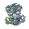
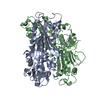
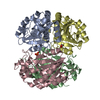
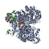
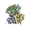
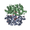
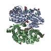
| Title | Biuret Hydrolase (BiuH) from Rhodococcus sp. Mel C169S Apo form | |||||||||
 Components Components | Biuret hydrolase | |||||||||
 Keywords Keywords | HYDROLASE / TrtA / BiuH / triuret / biuret / cysteine hydrolase / nitrogen | |||||||||
| Function / homology | : / Isochorismatase-like / Isochorismatase-like superfamily / Isochorismatase domain / hydrolase activity / Biuret hydrolase Function and homology information Function and homology information | |||||||||
| Biological species |  Rhodococcus sp. Mel (bacteria) Rhodococcus sp. Mel (bacteria) | |||||||||
| Method |  X-RAY DIFFRACTION / SYNCHROTRON / MOLECULAR REPLACEMENT / Resolution: 1.7 Å X-RAY DIFFRACTION / SYNCHROTRON / MOLECULAR REPLACEMENT / Resolution: 1.7 Å | |||||||||
 Authors Authors | Tassoulas, L.T. / Elias, M.H. / Wackett, L.P. | |||||||||
| Funding support |  United States, 2items United States, 2items
| |||||||||
 Citation Citation | Journal: J.Biol.Chem. / Year: 2020 Title: Discovery of an ultraspecific triuret hydrolase (TrtA) establishes the triuret biodegradation pathway. Authors: Tassoulas, L.J. / Elias, M.H. / Wackett, L.P. | |||||||||
| History |
|
- Structure visualization
| Structure viewer | Molecule: MolmilJmol/JSmol |
|---|
- Downloads & links
-Download
| PDBx/mmCIF format | 6xk1.cif.gz | 194 KB | Display | PDBx/mmCIF format |
|---|---|---|---|---|
| PDB format | pdb6xk1.ent.gz | 147.4 KB | Display | PDB format |
| PDBx/mmJSON format | 6xk1.json.gz | Tree view | PDBx/mmJSON format | |
| Others |  Other downloads Other downloads |
-Validation report
| Arichive directory | https://data.pdbj.org/pub/pdb/validation_reports/xk/6xk1ftp://data.pdbj.org/pub/pdb/validation_reports/xk/6xk1 | HTTPS FTP |
|---|
-Related structure data
| Related structure data |  6xixC  6xj4C  6xjeC 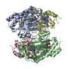 6xjmSC C: citing same article ( S: Starting model for refinement |
|---|---|
| Similar structure data |

-Links
 PDBj
PDBj- Assembly
| Deposited unit | 
| |||||||||||||||||||||
|---|---|---|---|---|---|---|---|---|---|---|---|---|---|---|---|---|---|---|---|---|---|---|
| 1 |
| |||||||||||||||||||||
| Unit cell |
| |||||||||||||||||||||
| Noncrystallographic symmetry (NCS) | NCS domain:
|
-Components
| #1: Protein | Mass: 26044.336 Da / Num. of mol.: 4 / Mutation: C169S Source method: isolated from a genetically manipulated source Source: (gene. exp.) Rhodococcus sp. Mel (bacteria) / Gene: biuH / Production host: #2: Water | ChemComp-HOH / |  Mass: 18.015 Da / Num. of mol.: 509 / Source method: isolated from a natural source / Formula: H2O Mass: 18.015 Da / Num. of mol.: 509 / Source method: isolated from a natural source / Formula: H2O |
|---|
-Experimental details
-Experiment
| Experiment | Method: X-RAY DIFFRACTION / Number of used crystals: 1 |
|---|
- Sample preparation
| Crystal | Density Matthews: 2.1 Å3/Da / Density % sol: 41.39 % |
|---|---|
| Crystal grow | Temperature: 291 K / Method: vapor diffusion, hanging drop / pH: 5.5 Details: 1uL 10mg/mL protein + 1uL 18% w/v PEG3350, 0.2M MgCl2, 1mM triuret pH 5.5 |
-Data collection
| Diffraction | Mean temperature: 100 K / Serial crystal experiment: N |
|---|---|
| Diffraction source | Source: SYNCHROTRON / Site: APS / Beamline: 23-ID-B / Wavelength: 0.99184 Å |
| Detector | Type: DECTRIS EIGER X 16M / Detector: PIXEL / Date: Mar 15, 2020 |
| Radiation | Monochromator: Double crystal cryo-cooled Si(111) / Protocol: SINGLE WAVELENGTH / Monochromatic (M) / Laue (L): M / Scattering type: x-ray |
| Radiation wavelength | Wavelength: 0.99184 Å / Relative weight: 1 |
| Reflection | Resolution: 1.7→82.66 Å / Num. obs: 186186 / % possible obs: 99.6 % / Redundancy: 3.85 % / Rmerge(I) obs: 0.077 / Net I/σ(I): 11.55 |
| Reflection shell | Resolution: 1.7→1.8 Å / Redundancy: 3.77 % / Rmerge(I) obs: 0.501 / Mean I/σ(I) obs: 2.96 / Num. unique obs: 29499 / % possible all: 99.9 |
- Processing
| Software |
| ||||||||||||||||||||||||||||||||||||||||||||||||||||||||||||||||||||||||||||||||||||||||||||||||||||||||||||||||||||||||||||||||||||||||||||||||||||||||||||||||||||||||||||||||||||
|---|---|---|---|---|---|---|---|---|---|---|---|---|---|---|---|---|---|---|---|---|---|---|---|---|---|---|---|---|---|---|---|---|---|---|---|---|---|---|---|---|---|---|---|---|---|---|---|---|---|---|---|---|---|---|---|---|---|---|---|---|---|---|---|---|---|---|---|---|---|---|---|---|---|---|---|---|---|---|---|---|---|---|---|---|---|---|---|---|---|---|---|---|---|---|---|---|---|---|---|---|---|---|---|---|---|---|---|---|---|---|---|---|---|---|---|---|---|---|---|---|---|---|---|---|---|---|---|---|---|---|---|---|---|---|---|---|---|---|---|---|---|---|---|---|---|---|---|---|---|---|---|---|---|---|---|---|---|---|---|---|---|---|---|---|---|---|---|---|---|---|---|---|---|---|---|---|---|---|---|---|---|
| Refinement | Method to determine structure: MOLECULAR REPLACEMENT Starting model: 6XJM Resolution: 1.7→56.837 Å / Cor.coef. Fo:Fc: 0.959 / Cor.coef. Fo:Fc free: 0.951 / WRfactor Rfree: 0.21 / WRfactor Rwork: 0.185 / SU B: 2.108 / SU ML: 0.069 / Average fsc free: 0.9342 / Average fsc work: 0.9422 / Cross valid method: THROUGHOUT / ESU R: 0.109 / ESU R Free: 0.102 Details: Hydrogens have been added in their riding positions
| ||||||||||||||||||||||||||||||||||||||||||||||||||||||||||||||||||||||||||||||||||||||||||||||||||||||||||||||||||||||||||||||||||||||||||||||||||||||||||||||||||||||||||||||||||||
| Solvent computation | Ion probe radii: 0.8 Å / Shrinkage radii: 0.8 Å / VDW probe radii: 1.2 Å / Solvent model: MASK BULK SOLVENT | ||||||||||||||||||||||||||||||||||||||||||||||||||||||||||||||||||||||||||||||||||||||||||||||||||||||||||||||||||||||||||||||||||||||||||||||||||||||||||||||||||||||||||||||||||||
| Displacement parameters | Biso mean: 18.828 Å2
| ||||||||||||||||||||||||||||||||||||||||||||||||||||||||||||||||||||||||||||||||||||||||||||||||||||||||||||||||||||||||||||||||||||||||||||||||||||||||||||||||||||||||||||||||||||
| Refinement step | Cycle: LAST / Resolution: 1.7→56.837 Å
| ||||||||||||||||||||||||||||||||||||||||||||||||||||||||||||||||||||||||||||||||||||||||||||||||||||||||||||||||||||||||||||||||||||||||||||||||||||||||||||||||||||||||||||||||||||
| Refine LS restraints |
| ||||||||||||||||||||||||||||||||||||||||||||||||||||||||||||||||||||||||||||||||||||||||||||||||||||||||||||||||||||||||||||||||||||||||||||||||||||||||||||||||||||||||||||||||||||
| LS refinement shell |
|
