 Movie
Movie Controller

[English] 日本語
 Yorodumi
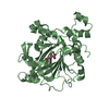
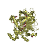
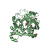
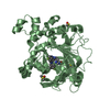
- PDB-6xjj: Structure of non-heme iron enzyme TropC: Radical tropolone biosyn... -
+ Open data

- Basic information
| Entry | Database: PDB / ID: 6xjj | ||||||
|---|---|---|---|---|---|---|---|
| Title | Structure of non-heme iron enzyme TropC: Radical tropolone biosynthesis | ||||||
 Components Components | 2-oxoglutarate-dependent dioxygenase tropC | ||||||
 Keywords Keywords | OXIDOREDUCTASE / Tropolone | ||||||
| Function / homology |  Function and homology information Function and homology informationsmall molecule biosynthetic process / Oxidoreductases; Acting on paired donors, with incorporation or reduction of molecular oxygen / dioxygenase activity / metal ion binding Similarity search - Function | ||||||
| Biological species | Talaromyces stipitatus | ||||||
| Method |  X-RAY DIFFRACTION / SYNCHROTRON / MOLECULAR REPLACEMENT / Resolution: 2.7 Å X-RAY DIFFRACTION / SYNCHROTRON / MOLECULAR REPLACEMENT / Resolution: 2.7 Å | ||||||
 Authors Authors | Mallik, L. / Doyon, T.J. / Narayan, A.R.H. / Koutmos, M. | ||||||
 Citation Citation | Journal: Chemrxiv / Year: 2020 Title: Radical Tropolone Biosynthesis Authors: Doyon, T.J. / Skinner, K. / Yang, D. / Mallik, L. / Wymore, T. / Koutmos, M. / Zimmerman, P.M. / Narayan, A. | ||||||
| History |
|
- Structure visualization
| Structure viewer | Molecule: MolmilJmol/JSmol |
|---|
- Downloads & links
-Download
| PDBx/mmCIF format | 6xjj.cif.gz | 120 KB | Display | PDBx/mmCIF format |
|---|---|---|---|---|
| PDB format | pdb6xjj.ent.gz | 94.1 KB | Display | PDB format |
| PDBx/mmJSON format | 6xjj.json.gz | Tree view | PDBx/mmJSON format | |
| Others |  Other downloads Other downloads |
-Validation report
| Arichive directory | https://data.pdbj.org/pub/pdb/validation_reports/xj/6xjjftp://data.pdbj.org/pub/pdb/validation_reports/xj/6xjj | HTTPS FTP |
|---|
-Related structure data
| Related structure data |  5c3qS S: Starting model for refinement |
|---|---|
| Similar structure data |


-Links
 PDBj
PDBj


- Assembly
| Deposited unit | 
| ||||||||
|---|---|---|---|---|---|---|---|---|---|
| 1 |
| ||||||||
| Unit cell |
|
-Components
| #1: Protein | Mass: 40063.512 Da / Num. of mol.: 1 Source method: isolated from a genetically manipulated source Source: (gene. exp.)  Talaromyces stipitatus (strain ATCC 10500 / CBS 375.48 / QM 6759 / NRRL 1006) (fungus) Talaromyces stipitatus (strain ATCC 10500 / CBS 375.48 / QM 6759 / NRRL 1006) (fungus)Strain: ATCC 10500 / CBS 375.48 / QM 6759 / NRRL 1006 / Gene: tropC, tsR5, TSTA_117800 / Production host:  References: UniProt: B8M9K5, Oxidoreductases; Acting on paired donors, with incorporation or reduction of molecular oxygen |
|---|---|
| #2: Chemical | ChemComp-FE /   Mass: 55.845 Da / Num. of mol.: 1 / Source method: obtained synthetically / Formula: Fe Mass: 55.845 Da / Num. of mol.: 1 / Source method: obtained synthetically / Formula: Fe |
| #3: Chemical | ChemComp-YT3 /   Mass: 88.906 Da / Num. of mol.: 1 / Source method: obtained synthetically / Formula: Y Mass: 88.906 Da / Num. of mol.: 1 / Source method: obtained synthetically / Formula: Y |
| #4: Chemical | ChemComp-ACT /   Mass: 59.044 Da / Num. of mol.: 1 / Source method: obtained synthetically / Formula: C2H3O2 Mass: 59.044 Da / Num. of mol.: 1 / Source method: obtained synthetically / Formula: C2H3O2 |
| #5: Water | ChemComp-HOH /  Mass: 18.015 Da / Num. of mol.: 5 / Source method: isolated from a natural source / Formula: H2O Mass: 18.015 Da / Num. of mol.: 5 / Source method: isolated from a natural source / Formula: H2O |
| Has ligand of interest | N |
-Experimental details
-Experiment
| Experiment | Method: X-RAY DIFFRACTION / Number of used crystals: 1 |
|---|
- Sample preparation
| Crystal | Density Matthews: 2.32 Å3/Da / Density % sol: 47 % |
|---|---|
| Crystal grow | Temperature: 277 K / Method: vapor diffusion, sitting drop / pH: 7.4 Details: 0.2 M Magnesium acetate, 20% PEG 8000, 0.01 M Yttrium (III) chloride hexahydrate |
-Data collection
| Diffraction | Mean temperature: 100 K / Serial crystal experiment: N |
|---|---|
| Diffraction source | Source: SYNCHROTRON / Site: APS  / Beamline: 21-ID-D / Wavelength: 1.0332 Å / Beamline: 21-ID-D / Wavelength: 1.0332 Å |
| Detector | Type: DECTRIS EIGER X 9M / Detector: PIXEL / Date: Apr 17, 2018 |
| Radiation | Monochromator: Si(111) / Protocol: SINGLE WAVELENGTH / Monochromatic (M) / Laue (L): M / Scattering type: x-ray |
| Radiation wavelength | Wavelength: 1.0332 Å / Relative weight: 1 |
| Reflection | Resolution: 2.7→48.07 Å / Num. obs: 17040 / % possible obs: 95.4 % / Redundancy: 5.9 % / Biso Wilson estimate: 64.9 Å2 / CC1/2: 0.99 / Rmerge(I) obs: 0.07 / Rpim(I) all: 0.034 / Rrim(I) all: 0.085 / Χ2: 1.01 / Net I/σ(I): 14.4 |
| Reflection shell | Resolution: 2.7→2.873 Å / Redundancy: 6 % / Rmerge(I) obs: 1.073 / Mean I/σ(I) obs: 1.7 / Num. unique obs: 2288 / CC1/2: 0.766 / Rpim(I) all: 0.72 / Rrim(I) all: 0.035 / Χ2: 1.04 / % possible all: 97.7 |
- Processing
| Software |
| |||||||||||||||||||||||||||||||||||||||||||||||||
|---|---|---|---|---|---|---|---|---|---|---|---|---|---|---|---|---|---|---|---|---|---|---|---|---|---|---|---|---|---|---|---|---|---|---|---|---|---|---|---|---|---|---|---|---|---|---|---|---|---|---|
| Refinement | Method to determine structure: MOLECULAR REPLACEMENT Starting model: 5C3Q Resolution: 2.7→48.07 Å / SU ML: 0.45 / Cross valid method: FREE R-VALUE / σ(F): 1.35 / Phase error: 29.59 / Stereochemistry target values: ML
| |||||||||||||||||||||||||||||||||||||||||||||||||
| Solvent computation | Shrinkage radii: 0.9 Å / VDW probe radii: 1.11 Å / Solvent model: FLAT BULK SOLVENT MODEL | |||||||||||||||||||||||||||||||||||||||||||||||||
| Displacement parameters | Biso mean: 64.9 Å2 | |||||||||||||||||||||||||||||||||||||||||||||||||
| Refinement step | Cycle: LAST / Resolution: 2.7→48.07 Å
| |||||||||||||||||||||||||||||||||||||||||||||||||
| Refine LS restraints |
| |||||||||||||||||||||||||||||||||||||||||||||||||
| LS refinement shell |
|
