 Movie
Movie Controller


+ Open data

- Basic information
| Entry | Database: PDB / ID: 6xj9 | ||||||
|---|---|---|---|---|---|---|---|
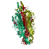
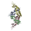
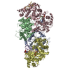
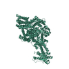
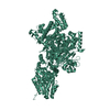
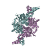
| Title | Structure of PfGH50B | ||||||
 Components Components | Agarase | ||||||
 Keywords Keywords | HYDROLASE / Glycoside hydrolase | ||||||
| Function / homology | Agarase, CBM-like domain / Agarase CBM like domain / Glycoside hydrolase, family 42, N-terminal / Beta-galactosidase / beta-galactosidase complex / beta-galactosidase activity / Glycoside hydrolase superfamily / carbohydrate metabolic process / Agarase Function and homology information Function and homology information | ||||||
| Biological species |  Pseudoalteromonas fuliginea (bacteria) Pseudoalteromonas fuliginea (bacteria) | ||||||
| Method |  X-RAY DIFFRACTION / SYNCHROTRON / MOLECULAR REPLACEMENT / molecular replacement / Resolution: 2 Å X-RAY DIFFRACTION / SYNCHROTRON / MOLECULAR REPLACEMENT / molecular replacement / Resolution: 2 Å | ||||||
 Authors Authors | Pluvinage, B. / Boraston, A.B. | ||||||
| Funding support |  Canada, 1items Canada, 1items
| ||||||
 Citation Citation | Journal: Acta Crystallogr.,Sect.F / Year: 2020 Title: The structure of PfGH50B, an agarase from the marine bacterium Pseudoalteromonas fuliginea PS47 Authors: Pluvinage, B. / Robb, C.S. / Jeffries, R. / Boraston, A.B. | ||||||
| History |
|
- Structure visualization
| Structure viewer | Molecule: MolmilJmol/JSmol |
|---|
- Downloads & links
-Download
| PDBx/mmCIF format | 6xj9.cif.gz | 291.6 KB | Display | PDBx/mmCIF format |
|---|---|---|---|---|
| PDB format | pdb6xj9.ent.gz | 230.2 KB | Display | PDB format |
| PDBx/mmJSON format | 6xj9.json.gz | Tree view | PDBx/mmJSON format | |
| Others |  Other downloads Other downloads |
-Validation report
| Summary document | 6xj9_validation.pdf.gz | 457.3 KB | Display | wwPDB validaton report |
|---|---|---|---|---|
| Full document | 6xj9_full_validation.pdf.gz | 469.3 KB | Display | |
| Data in XML | 6xj9_validation.xml.gz | 50.3 KB | Display | |
| Data in CIF | 6xj9_validation.cif.gz | 70.6 KB | Display | |
| Arichive directory | https://data.pdbj.org/pub/pdb/validation_reports/xj/6xj9ftp://data.pdbj.org/pub/pdb/validation_reports/xj/6xj9 | HTTPS FTP |
-Related structure data
| Related structure data |  4bq2S S: Starting model for refinement |
|---|---|
| Similar structure data |


-Links
 PDBj
PDBj- Assembly
| Deposited unit | 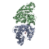
| ||||||||
|---|---|---|---|---|---|---|---|---|---|
| 1 |
| ||||||||
| Unit cell |
|
-Components
| #1: Protein | Mass: 86526.352 Da / Num. of mol.: 2 Source method: isolated from a genetically manipulated source Source: (gene. exp.) Pseudoalteromonas fuliginea (bacteria) / Gene: EUZ79_06745 / Plasmid: pET28a / Production host: #2: Chemical |   Mass: 62.068 Da / Num. of mol.: 2 / Source method: obtained synthetically / Formula: C2H6O2 Mass: 62.068 Da / Num. of mol.: 2 / Source method: obtained synthetically / Formula: C2H6O2#3: Water | ChemComp-HOH / |  Mass: 18.015 Da / Num. of mol.: 362 / Source method: isolated from a natural source / Formula: H2O Mass: 18.015 Da / Num. of mol.: 362 / Source method: isolated from a natural source / Formula: H2OHas ligand of interest | N | |
|---|
-Experimental details
-Experiment
| Experiment | Method: X-RAY DIFFRACTION / Number of used crystals: 1 |
|---|
- Sample preparation
| Crystal | Density Matthews: 2.18 Å3/Da / Density % sol: 43.6 % |
|---|---|
| Crystal grow | Temperature: 291 K / Method: vapor diffusion, hanging drop / pH: 6.5 / Details: 20% PEG 3350, 0.2M CaCl2, 0.1M MES |
-Data collection
| Diffraction | Mean temperature: 100 K / Serial crystal experiment: N |
|---|---|
| Diffraction source | Source: SYNCHROTRON / Site: CLSI / Beamline: 08B1-1 / Wavelength: 0.97932 Å |
| Detector | Type: RAYONIX MX-300 / Detector: CCD / Date: May 23, 2013 |
| Radiation | Protocol: SINGLE WAVELENGTH / Monochromatic (M) / Laue (L): M / Scattering type: x-ray |
| Radiation wavelength | Wavelength: 0.97932 Å / Relative weight: 1 |
| Reflection | Resolution: 2→29.98 Å / Num. obs: 99945 / % possible obs: 97.1 % / Redundancy: 6.7 % / CC1/2: 0.999 / Rmerge(I) obs: 0.058 / Rrim(I) all: 0.068 / Net I/σ(I): 18.3 |
| Reflection shell | Resolution: 2→2.03 Å / Redundancy: 4.7 % / Rmerge(I) obs: 0.525 / Mean I/σ(I) obs: 2.6 / Num. unique obs: 3724 / CC1/2: 0.723 / Rrim(I) all: 0.647 / % possible all: 74.9 |
-Phasing
| Phasing | Method: molecular replacement |
|---|
- Processing
| Software |
| |||||||||||||||||||||||||||||||||||||||||||||
|---|---|---|---|---|---|---|---|---|---|---|---|---|---|---|---|---|---|---|---|---|---|---|---|---|---|---|---|---|---|---|---|---|---|---|---|---|---|---|---|---|---|---|---|---|---|---|
| Refinement | Method to determine structure: MOLECULAR REPLACEMENT Starting model: 4BQ2 Resolution: 2→29.67 Å / Cor.coef. Fo:Fc: 0.934 / Cor.coef. Fo:Fc free: 0.906 / SU B: 5.47 / SU ML: 0.151 / Cross valid method: THROUGHOUT / σ(F): 0 / ESU R: 0.222 / ESU R Free: 0.192 / Stereochemistry target values: MAXIMUM LIKELIHOOD / Details: U VALUES : REFINED INDIVIDUALLY
| |||||||||||||||||||||||||||||||||||||||||||||
| Solvent computation | Ion probe radii: 0.8 Å / Shrinkage radii: 0.8 Å / VDW probe radii: 1.2 Å / Solvent model: MASK | |||||||||||||||||||||||||||||||||||||||||||||
| Displacement parameters | Biso max: 87.79 Å2 / Biso mean: 36.903 Å2 / Biso min: 13.5 Å2
| |||||||||||||||||||||||||||||||||||||||||||||
| Refinement step | Cycle: final / Resolution: 2→29.67 Å
| |||||||||||||||||||||||||||||||||||||||||||||
| Refine LS restraints |
| |||||||||||||||||||||||||||||||||||||||||||||
| LS refinement shell | Resolution: 2→2.052 Å / Rfactor Rfree error: 0 / Total num. of bins used: 20
|
