 Movie
Movie Controller

[English] 日本語
 Yorodumi
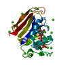
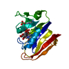
- PDB-6xg4: X-ray structure of Escherichia coli dihydrofolate reductase L28R ... -
+ Open data

- Basic information
| Entry | Database: PDB / ID: 6xg4 | |||||||||
|---|---|---|---|---|---|---|---|---|---|---|
| Title | X-ray structure of Escherichia coli dihydrofolate reductase L28R mutant in complex with trimethoprim | |||||||||
 Components Components | Dihydrofolate reductase | |||||||||
 Keywords Keywords | OXIDOREDUCTASE / DIHYDROFOLATE REDUCTASE / DHFR / MUTANT / COMPLEX / trimethoprim | |||||||||
| Function / homology |  Function and homology information Function and homology informationmethotrexate binding / dihydrofolic acid binding / 10-formyltetrahydrofolate biosynthetic process / response to methotrexate / folic acid biosynthetic process / folic acid binding / NADP+ binding / dihydrofolate metabolic process / dihydrofolate reductase / dihydrofolate reductase activity ...methotrexate binding / dihydrofolic acid binding / 10-formyltetrahydrofolate biosynthetic process / response to methotrexate / folic acid biosynthetic process / folic acid binding / NADP+ binding / dihydrofolate metabolic process / dihydrofolate reductase / dihydrofolate reductase activity / folic acid metabolic process / tetrahydrofolate biosynthetic process / NADPH binding / one-carbon metabolic process / NADP binding / response to xenobiotic stimulus / response to antibiotic / cytosol Similarity search - Function | |||||||||
| Biological species |  | |||||||||
| Method |  X-RAY DIFFRACTION / SYNCHROTRON / MOLECULAR REPLACEMENT / Resolution: 2.1 Å X-RAY DIFFRACTION / SYNCHROTRON / MOLECULAR REPLACEMENT / Resolution: 2.1 Å | |||||||||
 Authors Authors | Gaszek, I.K. / Manna, M.S. / Borek, D. / Toprak, E. | |||||||||
| Funding support |  United States, 2items United States, 2items
| |||||||||
 Citation Citation | Journal: Nat Commun / Year: 2021 Title: A trimethoprim derivative impedes antibiotic resistance evolution. Authors: Manna, M.S. / Tamer, Y.T. / Gaszek, I. / Poulides, N. / Ahmed, A. / Wang, X. / Toprak, F.C.R. / Woodard, D.R. / Koh, A.Y. / Williams, N.S. / Borek, D. / Atilgan, A.R. / Hulleman, J.D. / ...Authors: Manna, M.S. / Tamer, Y.T. / Gaszek, I. / Poulides, N. / Ahmed, A. / Wang, X. / Toprak, F.C.R. / Woodard, D.R. / Koh, A.Y. / Williams, N.S. / Borek, D. / Atilgan, A.R. / Hulleman, J.D. / Atilgan, C. / Tambar, U. / Toprak, E. | |||||||||
| History |
|
- Structure visualization
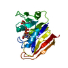
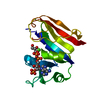
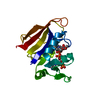
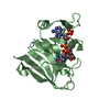
| Structure viewer | Molecule: MolmilJmol/JSmol |
|---|
- Downloads & links
-Download
| PDBx/mmCIF format | 6xg4.cif.gz | 89.4 KB | Display | PDBx/mmCIF format |
|---|---|---|---|---|
| PDB format | pdb6xg4.ent.gz | 66.3 KB | Display | PDB format |
| PDBx/mmJSON format | 6xg4.json.gz | Tree view | PDBx/mmJSON format | |
| Others |  Other downloads Other downloads |
-Validation report
| Arichive directory | https://data.pdbj.org/pub/pdb/validation_reports/xg/6xg4ftp://data.pdbj.org/pub/pdb/validation_reports/xg/6xg4 | HTTPS FTP |
|---|
-Related structure data


-Links
 PDBj
PDBj

- Assembly
| Deposited unit | 
| ||||||||
|---|---|---|---|---|---|---|---|---|---|
| 1 |
| ||||||||
| Unit cell |
| ||||||||
| Components on special symmetry positions |
|
-Components
-Protein , 1 types, 1 molecules A
| #1: Protein | Mass: 18892.260 Da / Num. of mol.: 1 / Mutation: L28R Source method: isolated from a genetically manipulated source Source: (gene. exp.) |
|---|
-Non-polymers , 5 types, 135 molecules 


| #2: Chemical | ChemComp-NDP /  Mass: 745.421 Da / Num. of mol.: 1 / Source method: obtained synthetically / Formula: C21H30N7O17P3 / Feature type: SUBJECT OF INVESTIGATION Mass: 745.421 Da / Num. of mol.: 1 / Source method: obtained synthetically / Formula: C21H30N7O17P3 / Feature type: SUBJECT OF INVESTIGATION |
|---|---|
| #3: Chemical | ChemComp-GOL /  Mass: 92.094 Da / Num. of mol.: 1 / Source method: obtained synthetically / Formula: C3H8O3 Mass: 92.094 Da / Num. of mol.: 1 / Source method: obtained synthetically / Formula: C3H8O3 |
| #4: Chemical | ChemComp-TOP /  Mass: 290.318 Da / Num. of mol.: 1 / Source method: obtained synthetically / Formula: C14H18N4O3 / Feature type: SUBJECT OF INVESTIGATION / Comment: antibiotic*YM Mass: 290.318 Da / Num. of mol.: 1 / Source method: obtained synthetically / Formula: C14H18N4O3 / Feature type: SUBJECT OF INVESTIGATION / Comment: antibiotic*YM |
| #5: Chemical | ChemComp-CL /  Mass: 35.453 Da / Num. of mol.: 1 / Source method: obtained synthetically / Formula: Cl Mass: 35.453 Da / Num. of mol.: 1 / Source method: obtained synthetically / Formula: Cl |
| #6: Water | ChemComp-HOH / Mass: 18.015 Da / Num. of mol.: 131 / Source method: isolated from a natural source / Formula: H2O |
-Details
| Has ligand of interest | Y |
|---|
-Experimental details
-Experiment
| Experiment | Method: X-RAY DIFFRACTION / Number of used crystals: 1 |
|---|
- Sample preparation
| Crystal | Density Matthews: 3.03 Å3/Da / Density % sol: 59.39 % |
|---|---|
| Crystal grow | Temperature: 293 K / Method: vapor diffusion, hanging drop / pH: 5.6 Details: 0.1 M sodium citrate tribasic dihydrate (pH 5.6), 0.15 M ammonium acetate and 17.5% or 20% PEG 4000; 10 mM NADPH, 2 mM TMP was incubated with the L28R variant of DHFR overnight at 293 K Temp details: RT |
-Data collection
| Diffraction | Mean temperature: 100 K / Serial crystal experiment: N |
|---|---|
| Diffraction source | Source: SYNCHROTRON / Site: APS / Beamline: 19-ID / Wavelength: 0.97919 Å |
| Detector | Type: DECTRIS PILATUS 6M / Detector: PIXEL / Date: Jul 12, 2018 |
| Radiation | Monochromator: Si(111) / Protocol: SINGLE WAVELENGTH / Monochromatic (M) / Laue (L): M / Scattering type: x-ray |
| Radiation wavelength | Wavelength: 0.97919 Å / Relative weight: 1 |
| Reflection | Resolution: 2.1→50 Å / Num. obs: 13880 / % possible obs: 99.9 % / Observed criterion σ(F): 0 / Observed criterion σ(I): -3 / Redundancy: 9.3 % / CC1/2: 1 / CC star: 1 / Rpim(I) all: 0.025 / Rrim(I) all: 0.076 / Net I/σ(I): 37.5 |
| Reflection shell | Resolution: 2.1→2.12 Å / Redundancy: 8.8 % / Mean I/σ(I) obs: 0.7 / Num. unique obs: 334 / CC1/2: 0.672 / CC star: 0.897 / Rpim(I) all: 3.012 / Rrim(I) all: 8.94 / % possible all: 100 |
- Processing
| Software |
| ||||||||||||||||||||||||||||||||||||||||||||||||||||||||||||
|---|---|---|---|---|---|---|---|---|---|---|---|---|---|---|---|---|---|---|---|---|---|---|---|---|---|---|---|---|---|---|---|---|---|---|---|---|---|---|---|---|---|---|---|---|---|---|---|---|---|---|---|---|---|---|---|---|---|---|---|---|---|
| Refinement | Method to determine structure: MOLECULAR REPLACEMENT Starting model: WT DHFR solved in the same space group Resolution: 2.1→37.37 Å / Cor.coef. Fo:Fc: 0.908 / SU B: 10.763 / SU ML: 0.16 / Cross valid method: THROUGHOUT / σ(F): 0 / ESU R: 0.369 / Stereochemistry target values: MAXIMUM LIKELIHOOD Details: HYDROGENS HAVE BEEN ADDED IN THE RIDING POSITIONS U VALUES : WITH TLS ADDED. Due to anisotropic lack of completeness, the model was first refined against all data, and the resulting model ...Details: HYDROGENS HAVE BEEN ADDED IN THE RIDING POSITIONS U VALUES : WITH TLS ADDED. Due to anisotropic lack of completeness, the model was first refined against all data, and the resulting model was refined twenty times in parallel, with each set of refinements using a different test set, until the twenty models converged. The R-free reported is the average of the twenty R-free values.
| ||||||||||||||||||||||||||||||||||||||||||||||||||||||||||||
| Solvent computation | Ion probe radii: 0.8 Å / Shrinkage radii: 0.8 Å / VDW probe radii: 1.2 Å / Solvent model: MASK | ||||||||||||||||||||||||||||||||||||||||||||||||||||||||||||
| Displacement parameters | Biso max: 88.96 Å2 / Biso mean: 35.198 Å2 / Biso min: 13.54 Å2
| ||||||||||||||||||||||||||||||||||||||||||||||||||||||||||||
| Refinement step | Cycle: final / Resolution: 2.1→37.37 Å
| ||||||||||||||||||||||||||||||||||||||||||||||||||||||||||||
| Refine LS restraints |
| ||||||||||||||||||||||||||||||||||||||||||||||||||||||||||||
| LS refinement shell | Resolution: 2.103→2.157 Å / Rfactor Rfree error: 0 / Total num. of bins used: 20
| ||||||||||||||||||||||||||||||||||||||||||||||||||||||||||||
| Refinement TLS params. | Method: refined / Origin x: -11.393 Å / Origin y: 22.809 Å / Origin z: 16.417 Å
| ||||||||||||||||||||||||||||||||||||||||||||||||||||||||||||
| Refinement TLS group |
|
