 Movie
Movie Controller

[English] 日本語
 Yorodumi
- PDB-6xfk: Crystal structure of the type III secretion system pilotin-secret... -
+ Open data

- Basic information
| Entry | Database: PDB / ID: 6xfk | |||||||||
|---|---|---|---|---|---|---|---|---|---|---|
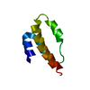
| Title | Crystal structure of the type III secretion system pilotin-secretin complex InvH-InvG | |||||||||
 Components Components |
| |||||||||
 Keywords Keywords | PROTEIN TRANSPORT / pilotin / secretin / chaperone / type III secretion | |||||||||
| Function / homology |  Function and homology information Function and homology informationtype III protein secretion system complex / type II protein secretion system complex / protein secretion by the type III secretion system / protein secretion / cell outer membrane / identical protein binding Similarity search - Function | |||||||||
| Biological species |  Salmonella typhimurium (bacteria) Salmonella typhimurium (bacteria) | |||||||||
| Method |  X-RAY DIFFRACTION / SYNCHROTRON / MOLECULAR REPLACEMENT / Resolution: 1.85 Å X-RAY DIFFRACTION / SYNCHROTRON / MOLECULAR REPLACEMENT / Resolution: 1.85 Å | |||||||||
 Authors Authors | Majewski, D.D. / Okon, M. / Heinkel, F. / Robb, C.S. / Vuckovic, M. / McIntosh, L.P. / Strynadka, N.C.J. | |||||||||
| Funding support |  Canada, 2items Canada, 2items
| |||||||||
 Citation Citation | Journal: Structure / Year: 2021 Title: Characterization of the Pilotin-Secretin Complex from the Salmonella enterica Type III Secretion System Using Hybrid Structural Methods. Authors: Majewski, D.D. / Okon, M. / Heinkel, F. / Robb, C.S. / Vuckovic, M. / McIntosh, L.P. / Strynadka, N.C.J. | |||||||||
| History |
|
- Structure visualization
| Structure viewer | Molecule: MolmilJmol/JSmol |
|---|
- Downloads & links
-Download
| PDBx/mmCIF format | 6xfk.cif.gz | 49.5 KB | Display | PDBx/mmCIF format |
|---|---|---|---|---|
| PDB format | pdb6xfk.ent.gz | 33.9 KB | Display | PDB format |
| PDBx/mmJSON format | 6xfk.json.gz | Tree view | PDBx/mmJSON format | |
| Others |  Other downloads Other downloads |
-Validation report
| Arichive directory | https://data.pdbj.org/pub/pdb/validation_reports/xf/6xfkftp://data.pdbj.org/pub/pdb/validation_reports/xf/6xfk | HTTPS FTP |
|---|
-Related structure data
| Related structure data |  6xfjSC  6xflC S: Starting model for refinement C: citing same article ( |
|---|---|
| Similar structure data |


-Links
 PDBj
PDBj


- Assembly
| Deposited unit | 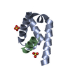
| |||||||||
|---|---|---|---|---|---|---|---|---|---|---|
| 1 |
| |||||||||
| Unit cell |
| |||||||||
| Components on special symmetry positions |
|
-Components
| #1: Protein | Mass: 8090.240 Da / Num. of mol.: 1 Source method: isolated from a genetically manipulated source Source: (gene. exp.) Salmonella typhimurium (strain LT2 / SGSC1412 / ATCC 700720) (bacteria)Strain: LT2 / SGSC1412 / ATCC 700720 / Gene: invH, sctG, STM2900 / Production host: | ||||||
|---|---|---|---|---|---|---|---|
| #2: Protein/peptide | Mass: 2023.296 Da / Num. of mol.: 1 / Source method: obtained synthetically Source: (synth.) Salmonella typhimurium (strain LT2 / SGSC1412 / ATCC 700720) (bacteria)References: UniProt: P35672 | ||||||
| #3: Chemical |   Mass: 96.063 Da / Num. of mol.: 2 / Source method: obtained synthetically / Formula: SO4 Mass: 96.063 Da / Num. of mol.: 2 / Source method: obtained synthetically / Formula: SO4#4: Water | ChemComp-HOH / |  Mass: 18.015 Da / Num. of mol.: 69 / Source method: isolated from a natural source / Formula: H2O Mass: 18.015 Da / Num. of mol.: 69 / Source method: isolated from a natural source / Formula: H2OHas ligand of interest | N | Has protein modification | Y | |
-Experimental details
-Experiment
| Experiment | Method: X-RAY DIFFRACTION / Number of used crystals: 1 |
|---|
- Sample preparation
| Crystal | Density Matthews: 2.09 Å3/Da / Density % sol: 41.01 % |
|---|---|
| Crystal grow | Temperature: 291 K / Method: vapor diffusion, hanging drop / pH: 4 Details: 0.5 M lithium sulfate, 8.3% PEG 6000, 100 mM sodium acetate |
-Data collection
| Diffraction | Mean temperature: 100 K / Serial crystal experiment: N |
|---|---|
| Diffraction source | Source: SYNCHROTRON / Site: ALS  / Beamline: 5.0.2 / Wavelength: 1 Å / Beamline: 5.0.2 / Wavelength: 1 Å |
| Detector | Type: DECTRIS PILATUS3 6M / Detector: PIXEL / Date: Mar 7, 2020 |
| Radiation | Protocol: SINGLE WAVELENGTH / Monochromatic (M) / Laue (L): M / Scattering type: x-ray |
| Radiation wavelength | Wavelength: 1 Å / Relative weight: 1 |
| Reflection | Resolution: 1.85→36.93 Å / Num. obs: 8226 / % possible obs: 99.5 % / Redundancy: 16.5 % / CC1/2: 1 / Net I/σ(I): 16.8 |
| Reflection shell | Resolution: 1.85→1.92 Å / Num. unique obs: 788 / CC1/2: 0.908 |
- Processing
| Software |
| |||||||||||||||||||||||||||||||||||||||||||||||||||||||||||||||||||||||||||
|---|---|---|---|---|---|---|---|---|---|---|---|---|---|---|---|---|---|---|---|---|---|---|---|---|---|---|---|---|---|---|---|---|---|---|---|---|---|---|---|---|---|---|---|---|---|---|---|---|---|---|---|---|---|---|---|---|---|---|---|---|---|---|---|---|---|---|---|---|---|---|---|---|---|---|---|---|
| Refinement | Method to determine structure: MOLECULAR REPLACEMENT Starting model: 6XFJ Resolution: 1.85→36.93 Å / Cor.coef. Fo:Fc: 0.948 / Cor.coef. Fo:Fc free: 0.943 / SU B: 6.794 / SU ML: 0.101 / Cross valid method: THROUGHOUT / σ(F): 0 / ESU R: 0.155 / ESU R Free: 0.14 / Stereochemistry target values: MAXIMUM LIKELIHOOD Details: HYDROGENS HAVE BEEN ADDED IN THE RIDING POSITIONS U VALUES : WITH TLS ADDED
| |||||||||||||||||||||||||||||||||||||||||||||||||||||||||||||||||||||||||||
| Solvent computation | Ion probe radii: 0.8 Å / Shrinkage radii: 0.8 Å / VDW probe radii: 1.2 Å / Solvent model: MASK | |||||||||||||||||||||||||||||||||||||||||||||||||||||||||||||||||||||||||||
| Displacement parameters | Biso max: 139.6 Å2 / Biso mean: 43.558 Å2 / Biso min: 27.51 Å2
| |||||||||||||||||||||||||||||||||||||||||||||||||||||||||||||||||||||||||||
| Refinement step | Cycle: final / Resolution: 1.85→36.93 Å
| |||||||||||||||||||||||||||||||||||||||||||||||||||||||||||||||||||||||||||
| Refine LS restraints |
| |||||||||||||||||||||||||||||||||||||||||||||||||||||||||||||||||||||||||||
| LS refinement shell | Resolution: 1.85→1.898 Å / Rfactor Rfree error: 0 / Total num. of bins used: 20
| |||||||||||||||||||||||||||||||||||||||||||||||||||||||||||||||||||||||||||
| Refinement TLS params. | Method: refined / Refine-ID: X-RAY DIFFRACTION
| |||||||||||||||||||||||||||||||||||||||||||||||||||||||||||||||||||||||||||
| Refinement TLS group |
|
