[English] 日本語
 Yorodumi
- PDB-6xbd: Cryo-EM structure of MlaFEDB in nanodiscs with phospholipid substrates -
+ Open data
- Basic information
| Entry | Database: PDB / ID: 6xbd | |||||||||||||||||||||||||||||||||||||||
|---|---|---|---|---|---|---|---|---|---|---|---|---|---|---|---|---|---|---|---|---|---|---|---|---|---|---|---|---|---|---|---|---|---|---|---|---|---|---|---|---|
| Title | Cryo-EM structure of MlaFEDB in nanodiscs with phospholipid substrates | |||||||||||||||||||||||||||||||||||||||
 Components Components |
| |||||||||||||||||||||||||||||||||||||||
 Keywords Keywords | LIPID TRANSPORT / bacterial cell envelope / mla pathway / MCE | |||||||||||||||||||||||||||||||||||||||
| Function / homology |  Function and homology information Function and homology informationHydrolases; Acting on acid anhydrides; Acting on acid anhydrides to catalyse transmembrane movement of substances / phospholipid transport / ATPase-coupled transmembrane transporter activity / ATP-binding cassette (ABC) transporter complex / hydrolase activity / ATP binding Similarity search - Function | |||||||||||||||||||||||||||||||||||||||
| Biological species |    Homo sapiens (human) Homo sapiens (human) | |||||||||||||||||||||||||||||||||||||||
| Method | ELECTRON MICROSCOPY / single particle reconstruction / cryo EM / Resolution: 3.05 Å | |||||||||||||||||||||||||||||||||||||||
 Authors Authors | Coudray, N. / Isom, G.L. / MacRae, M.R. / Saiduddin, M. / Ekiert, D.C. / Bhabha, G. | |||||||||||||||||||||||||||||||||||||||
| Funding support |  United States, 12items United States, 12items
| |||||||||||||||||||||||||||||||||||||||
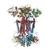
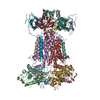
 Citation Citation |  Journal: Elife / Year: 2020 Journal: Elife / Year: 2020Title: Structure of bacterial phospholipid transporter MlaFEDB with substrate bound. Authors: Nicolas Coudray / Georgia L Isom / Mark R MacRae / Mariyah N Saiduddin / Gira Bhabha / Damian C Ekiert /  Abstract: In double-membraned bacteria, phospholipid transport across the cell envelope is critical to maintain the outer membrane barrier, which plays a key role in virulence and antibiotic resistance. An MCE ...In double-membraned bacteria, phospholipid transport across the cell envelope is critical to maintain the outer membrane barrier, which plays a key role in virulence and antibiotic resistance. An MCE transport system called Mla has been implicated in phospholipid trafficking and outer membrane integrity, and includes an ABC transporter, MlaFEDB. The transmembrane subunit, MlaE, has minimal sequence similarity to other transporters, and the structure of the entire inner-membrane MlaFEDB complex remains unknown. Here, we report the cryo-EM structure of MlaFEDB at 3.05 Å resolution, revealing distant relationships to the LPS and MacAB transporters, as well as the eukaryotic ABCA/ABCG families. A continuous transport pathway extends from the MlaE substrate-binding site, through the channel of MlaD, and into the periplasm. Unexpectedly, two phospholipids are bound to MlaFEDB, suggesting that multiple lipid substrates may be transported each cycle. Our structure provides mechanistic insight into substrate recognition and transport by MlaFEDB. | |||||||||||||||||||||||||||||||||||||||
| History |
|
- Structure visualization
| Movie |
 Movie viewer Movie viewer |
|---|---|
| Structure viewer | Molecule:  Molmil Molmil Jmol/JSmol Jmol/JSmol |
- Downloads & links
- Download
| PDBx/mmCIF format |  6xbd.cif.gz 6xbd.cif.gz | 388.9 KB | Display |  PDBx/mmCIF format PDBx/mmCIF format |
|---|---|---|---|---|
| PDB format |  pdb6xbd.ent.gz pdb6xbd.ent.gz | 316.3 KB | Display |  PDB format PDB format |
| PDBx/mmJSON format |  6xbd.json.gz 6xbd.json.gz | Tree view |  PDBx/mmJSON format PDBx/mmJSON format | |
| Others |  Other downloads Other downloads |
-Validation report
| Summary document |  6xbd_validation.pdf.gz 6xbd_validation.pdf.gz | 1 MB | Display |  wwPDB validaton report wwPDB validaton report |
|---|---|---|---|---|
| Full document |  6xbd_full_validation.pdf.gz 6xbd_full_validation.pdf.gz | 1.1 MB | Display | |
| Data in XML |  6xbd_validation.xml.gz 6xbd_validation.xml.gz | 60.7 KB | Display | |
| Data in CIF |  6xbd_validation.cif.gz 6xbd_validation.cif.gz | 93.3 KB | Display | |
| Arichive directory |  https://data.pdbj.org/pub/pdb/validation_reports/xb/6xbd https://data.pdbj.org/pub/pdb/validation_reports/xb/6xbd ftp://data.pdbj.org/pub/pdb/validation_reports/xb/6xbd ftp://data.pdbj.org/pub/pdb/validation_reports/xb/6xbd | HTTPS FTP |
-Related structure data
| Related structure data |  22116MC M: map data used to model this data C: citing same article ( |
|---|---|
| Similar structure data | |
| EM raw data |  EMPIAR-10536 (Title: Single particle cryo-EM dataset for MlaFEDB from E. coli in nanodisc EMPIAR-10536 (Title: Single particle cryo-EM dataset for MlaFEDB from E. coli in nanodiscData size: 5.2 TB Data #1: Unaligned multi-frame micrographs of MlaFEDB complex [micrographs - multiframe] Data #2: Polished particles (using Relion 3.1) [picked particles - single frame - processed]) |
- Links
Links
- Assembly
| Deposited unit | 
|
|---|---|
| 1 |
|
- Components
-Phospholipid ... , 4 types, 12 molecules ABCDEFGHIJKL
| #1: Protein | Mass: 21937.674 Da / Num. of mol.: 6 Source method: isolated from a genetically manipulated source Source: (gene. exp.)   #2: Protein | Mass: 27885.162 Da / Num. of mol.: 2 Source method: isolated from a genetically manipulated source Source: (gene. exp.)   #3: Protein | Mass: 29128.801 Da / Num. of mol.: 2 Source method: isolated from a genetically manipulated source Source: (gene. exp.)   References: UniProt: H4UPQ0, Hydrolases; Acting on acid anhydrides; Acting on acid anhydrides to catalyse transmembrane movement of substances #4: Protein | Mass: 10690.313 Da / Num. of mol.: 2 Source method: isolated from a genetically manipulated source Source: (gene. exp.)   |
|---|
-Protein / Non-polymers , 2 types, 4 molecules MN

| #5: Protein | Mass: 13975.214 Da / Num. of mol.: 2 Source method: isolated from a genetically manipulated source Source: (gene. exp.)  Homo sapiens (human) / Production host: Homo sapiens (human) / Production host:  #6: Chemical | |
|---|
-Details
| Compound details | Molecule-4 was modeled as a poly-ala chain. The register of the model with the sequence is unknown |
|---|---|
| Has ligand of interest | Y |
| Sequence details | sequence of entity-4 MSP1D1 ...sequence of entity-4 MSP1D1 STFSKLREQL |
-Experimental details
-Experiment
| Experiment | Method: ELECTRON MICROSCOPY |
|---|---|
| EM experiment | Aggregation state: PARTICLE / 3D reconstruction method: single particle reconstruction |
- Sample preparation
| Component | Name: MlaFEDB complex with two bound phospholipid substrates in MSP1D1 nanodisc Type: COMPLEX / Entity ID: #1-#5 / Source: RECOMBINANT |
|---|---|
| Molecular weight | Value: 0.266 MDa / Experimental value: NO |
| Source (natural) | Organism:  |
| Source (recombinant) | Organism:  |
| Buffer solution | pH: 7.4 |
| Specimen | Conc.: 0.95 mg/ml / Embedding applied: NO / Shadowing applied: NO / Staining applied: NO / Vitrification applied: YES |
| Specimen support | Grid mesh size: 400 divisions/in. / Grid type: Quantifoil R1.2/1.3 |
| Vitrification | Instrument: FEI VITROBOT MARK IV / Cryogen name: ETHANE |
- Electron microscopy imaging
| Experimental equipment |  Model: Titan Krios / Image courtesy: FEI Company |
|---|---|
| Microscopy | Model: FEI TITAN KRIOS |
| Electron gun | Electron source:  FIELD EMISSION GUN / Accelerating voltage: 300 kV / Illumination mode: FLOOD BEAM FIELD EMISSION GUN / Accelerating voltage: 300 kV / Illumination mode: FLOOD BEAM |
| Electron lens | Mode: BRIGHT FIELD / Nominal magnification: 29000 X / Cs: 2.7 mm |
| Specimen holder | Specimen holder model: FEI TITAN KRIOS AUTOGRID HOLDER |
| Image recording | Average exposure time: 6 sec. / Electron dose: 71 e/Å2 / Detector mode: SUPER-RESOLUTION / Film or detector model: GATAN K2 SUMMIT (4k x 4k) / Num. of grids imaged: 1 / Num. of real images: 3212 |
| Image scans | Movie frames/image: 30 / Used frames/image: 1-30 |
- Processing
| Software | Name: PHENIX / Version: 1.16_3549: / Classification: refinement | |||||||||||||||||||||||||||||||||||||||||||||||||||||||||||||||||
|---|---|---|---|---|---|---|---|---|---|---|---|---|---|---|---|---|---|---|---|---|---|---|---|---|---|---|---|---|---|---|---|---|---|---|---|---|---|---|---|---|---|---|---|---|---|---|---|---|---|---|---|---|---|---|---|---|---|---|---|---|---|---|---|---|---|---|
| EM software |
| |||||||||||||||||||||||||||||||||||||||||||||||||||||||||||||||||
| CTF correction | Type: PHASE FLIPPING AND AMPLITUDE CORRECTION | |||||||||||||||||||||||||||||||||||||||||||||||||||||||||||||||||
| Particle selection | Num. of particles selected: 1283606 | |||||||||||||||||||||||||||||||||||||||||||||||||||||||||||||||||
| Symmetry | Point symmetry: C1 (asymmetric) | |||||||||||||||||||||||||||||||||||||||||||||||||||||||||||||||||
| 3D reconstruction | Resolution: 3.05 Å / Resolution method: FSC 0.143 CUT-OFF / Num. of particles: 731205 / Algorithm: BACK PROJECTION / Num. of class averages: 1 / Symmetry type: POINT | |||||||||||||||||||||||||||||||||||||||||||||||||||||||||||||||||
| Atomic model building | Protocol: OTHER / Space: REAL | |||||||||||||||||||||||||||||||||||||||||||||||||||||||||||||||||
| Atomic model building | 3D fitting-ID: 1 / Source name: PDB / Type: experimental model
| |||||||||||||||||||||||||||||||||||||||||||||||||||||||||||||||||
| Refine LS restraints |
|
 Movie
Movie Controller


 PDBj
PDBj


