 Movie
Movie Controller

[English] 日本語
 Yorodumi
- PDB-6x7x: mannose-bound structure of Marinomonas primoryensis PA14 carbohyd... -
+ Open data

- Basic information
| Entry | Database: PDB / ID: 6x7x | ||||||
|---|---|---|---|---|---|---|---|
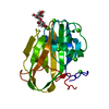
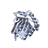
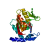
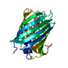
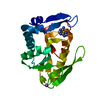
| Title | mannose-bound structure of Marinomonas primoryensis PA14 carbohydrate-binding domain | ||||||
 Components Components | Antifreeze protein | ||||||
 Keywords Keywords | SUGAR BINDING PROTEIN / mannose-MpPA14 complex / PA14 domain / carbohydrate-binding protein | ||||||
| Function / homology |  Function and homology information Function and homology information | ||||||
| Biological species |  Marinomonas primoryensis (bacteria) Marinomonas primoryensis (bacteria) | ||||||
| Method |  X-RAY DIFFRACTION / SYNCHROTRON / MOLECULAR REPLACEMENT / Resolution: 1.3 Å X-RAY DIFFRACTION / SYNCHROTRON / MOLECULAR REPLACEMENT / Resolution: 1.3 Å | ||||||
 Authors Authors | Guo, S. / Davies, P.L. | ||||||
| Funding support | 1items
| ||||||
 Citation Citation | Journal: Mbio / Year: 2021 Title: Structural Basis of Ligand Selectivity by a Bacterial Adhesin Lectin Involved in Multispecies Biofilm Formation. Authors: Guo, S. / Vance, T.D.R. / Zahiri, H. / Eves, R. / Stevens, C. / Hehemann, J.H. / Vidal-Melgosa, S. / Davies, P.L. #1: Journal: To Be PublishedTitle: Molecular basis for a bacterial adhesins sugar-binding module Authors: Guo, S. | ||||||
| History |
|
- Structure visualization
| Structure viewer | Molecule: MolmilJmol/JSmol |
|---|
- Downloads & links
-Download
| PDBx/mmCIF format | 6x7x.cif.gz | 115.9 KB | Display | PDBx/mmCIF format |
|---|---|---|---|---|
| PDB format | pdb6x7x.ent.gz | 76.3 KB | Display | PDB format |
| PDBx/mmJSON format | 6x7x.json.gz | Tree view | PDBx/mmJSON format | |
| Others |  Other downloads Other downloads |
-Validation report
| Arichive directory | https://data.pdbj.org/pub/pdb/validation_reports/x7/6x7xftp://data.pdbj.org/pub/pdb/validation_reports/x7/6x7x | HTTPS FTP |
|---|
-Related structure data
| Related structure data | 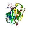 6x7jC  6x7tC  6x7yC  6x7zC  6x8aC  6x8dC  6x8yC  6x95C  6x9mC  6x9pC  6xa5C  6xacC  6xaqC 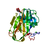 5j6yS S: Starting model for refinement C: citing same article ( |
|---|---|
| Similar structure data |


-Links
 PDBj
PDBj


- Assembly
| Deposited unit | 
| ||||||||||||
|---|---|---|---|---|---|---|---|---|---|---|---|---|---|
| 1 |
| ||||||||||||
| Unit cell |
|
-Components
-Protein , 1 types, 1 molecules A
| #1: Protein | Mass: 20249.479 Da / Num. of mol.: 1 / Fragment: carbohydrate-binding domain Source method: isolated from a genetically manipulated source Source: (gene. exp.) Marinomonas primoryensis (bacteria) / Production host: |
|---|
-Sugars , 2 types, 2 molecules 

| #4: Sugar | ChemComp-MAN /  Type: D-saccharide, alpha linking / Mass: 180.156 Da / Num. of mol.: 1 / Source method: obtained synthetically / Formula: C6H12O6 / Feature type: SUBJECT OF INVESTIGATION Type: D-saccharide, alpha linking / Mass: 180.156 Da / Num. of mol.: 1 / Source method: obtained synthetically / Formula: C6H12O6 / Feature type: SUBJECT OF INVESTIGATION |
|---|---|
| #5: Sugar | ChemComp-BMA /  Type: D-saccharide, beta linking / Mass: 180.156 Da / Num. of mol.: 1 / Source method: obtained synthetically / Formula: C6H12O6 / Feature type: SUBJECT OF INVESTIGATION Type: D-saccharide, beta linking / Mass: 180.156 Da / Num. of mol.: 1 / Source method: obtained synthetically / Formula: C6H12O6 / Feature type: SUBJECT OF INVESTIGATION |
-Non-polymers , 3 types, 275 molecules 


| #2: Chemical | ChemComp-CA /  Mass: 40.078 Da / Num. of mol.: 9 / Source method: obtained synthetically / Formula: Ca Mass: 40.078 Da / Num. of mol.: 9 / Source method: obtained synthetically / Formula: Ca#3: Chemical |  Mass: 62.068 Da / Num. of mol.: 2 / Source method: obtained synthetically / Formula: C2H6O2 Mass: 62.068 Da / Num. of mol.: 2 / Source method: obtained synthetically / Formula: C2H6O2#6: Water | ChemComp-HOH / | Mass: 18.015 Da / Num. of mol.: 264 / Source method: isolated from a natural source / Formula: H2O |
|---|
-Details
| Has ligand of interest | Y |
|---|
-Experimental details
-Experiment
| Experiment | Method: X-RAY DIFFRACTION / Number of used crystals: 1 |
|---|
- Sample preparation
| Crystal | Density Matthews: 2.26 Å3/Da / Density % sol: 45.47 % |
|---|---|
| Crystal grow | Temperature: 298 K / Method: microbatch / pH: 7 Details: 0.2 M calcium chloride, 0.1 M HEPES (pH 7), 20% (v/v) polyethylene glycol 3350 and ~30 % (w/v) mannose |
-Data collection
| Diffraction | Mean temperature: 93 K / Serial crystal experiment: N |
|---|---|
| Diffraction source | Source: SYNCHROTRON / Site: APS  / Beamline: 19-ID / Wavelength: 1.03318 Å / Beamline: 19-ID / Wavelength: 1.03318 Å |
| Detector | Type: MAR CCD 130 mm / Detector: CCD / Date: Apr 1, 2016 |
| Radiation | Protocol: SINGLE WAVELENGTH / Monochromatic (M) / Laue (L): M / Scattering type: x-ray |
| Radiation wavelength | Wavelength: 1.03318 Å / Relative weight: 1 |
| Reflection | Resolution: 1.26→39.56 Å / Num. obs: 48977 / % possible obs: 98.66 % / Redundancy: 7.7 % / Biso Wilson estimate: 5.2 Å2 / Rmerge(I) obs: 0.302 / Net I/σ(I): 4.2 |
| Reflection shell | Resolution: 1.26→1.31 Å / Rmerge(I) obs: 0.661 / Num. unique obs: 4287 |
- Processing
| Software | Name: PHENIX / Version: 1.17.1_3660 / Classification: refinement | ||||||||||||||||||||||||||||||||||||||||||||||||||||||||||||||||||||||||||||||||||||||||||||||||||||||||||||||||||||||||||||||
|---|---|---|---|---|---|---|---|---|---|---|---|---|---|---|---|---|---|---|---|---|---|---|---|---|---|---|---|---|---|---|---|---|---|---|---|---|---|---|---|---|---|---|---|---|---|---|---|---|---|---|---|---|---|---|---|---|---|---|---|---|---|---|---|---|---|---|---|---|---|---|---|---|---|---|---|---|---|---|---|---|---|---|---|---|---|---|---|---|---|---|---|---|---|---|---|---|---|---|---|---|---|---|---|---|---|---|---|---|---|---|---|---|---|---|---|---|---|---|---|---|---|---|---|---|---|---|---|
| Refinement | Method to determine structure: MOLECULAR REPLACEMENT Starting model: 5j6Y Resolution: 1.3→39.56 Å / SU ML: 0.1052 / Cross valid method: FREE R-VALUE / σ(F): 1.34 / Phase error: 16.1439 Stereochemistry target values: GeoStd + Monomer Library + CDL v1.2
| ||||||||||||||||||||||||||||||||||||||||||||||||||||||||||||||||||||||||||||||||||||||||||||||||||||||||||||||||||||||||||||||
| Solvent computation | Shrinkage radii: 0.9 Å / VDW probe radii: 1.11 Å / Solvent model: FLAT BULK SOLVENT MODEL | ||||||||||||||||||||||||||||||||||||||||||||||||||||||||||||||||||||||||||||||||||||||||||||||||||||||||||||||||||||||||||||||
| Displacement parameters | Biso mean: 10.06 Å2 | ||||||||||||||||||||||||||||||||||||||||||||||||||||||||||||||||||||||||||||||||||||||||||||||||||||||||||||||||||||||||||||||
| Refinement step | Cycle: LAST / Resolution: 1.3→39.56 Å
| ||||||||||||||||||||||||||||||||||||||||||||||||||||||||||||||||||||||||||||||||||||||||||||||||||||||||||||||||||||||||||||||
| Refine LS restraints |
| ||||||||||||||||||||||||||||||||||||||||||||||||||||||||||||||||||||||||||||||||||||||||||||||||||||||||||||||||||||||||||||||
| LS refinement shell |
|
