 Movie
Movie Controller

[English] 日本語
 Yorodumi
- PDB-6x6m: Peptide-bound structure of Marinomonas primoryensis peptide-bindi... -
+ Open data

- Basic information
| Entry | Database: PDB / ID: 6x6m | ||||||
|---|---|---|---|---|---|---|---|
| Title | Peptide-bound structure of Marinomonas primoryensis peptide-binding domain | ||||||
 Components Components |
| ||||||
 Keywords Keywords | PROTEIN BINDING / peptide-binding domain / protein-binding domain | ||||||
| Function / homology |  Function and homology information Function and homology information | ||||||
| Biological species |  Marinomonas primoryensis (bacteria) Marinomonas primoryensis (bacteria)synthetic construct (others) | ||||||
| Method |  X-RAY DIFFRACTION / SYNCHROTRON / MOLECULAR REPLACEMENT / Resolution: 1.9 Å X-RAY DIFFRACTION / SYNCHROTRON / MOLECULAR REPLACEMENT / Resolution: 1.9 Å | ||||||
 Authors Authors | Guo, S. / Davies, P.L. | ||||||
| Funding support | 1items
| ||||||
 Citation Citation | Journal: Cell Rep / Year: 2021 Title: Molecular basis for inhibition of adhesin-mediated bacterial-host interactions through a peptide-binding domain. Authors: Guo, S. / Zahiri, H. / Stevens, C. / Spaanderman, D.C. / Milroy, L.G. / Ottmann, C. / Brunsveld, L. / Voets, I.K. / Davies, P.L. | ||||||
| History |
|
- Structure visualization
| Structure viewer | Molecule: MolmilJmol/JSmol |
|---|
- Downloads & links
-Download
| PDBx/mmCIF format | 6x6m.cif.gz | 348.7 KB | Display | PDBx/mmCIF format |
|---|---|---|---|---|
| PDB format | pdb6x6m.ent.gz | 250.7 KB | Display | PDB format |
| PDBx/mmJSON format | 6x6m.json.gz | Tree view | PDBx/mmJSON format | |
| Others |  Other downloads Other downloads |
-Validation report
| Arichive directory | https://data.pdbj.org/pub/pdb/validation_reports/x6/6x6mftp://data.pdbj.org/pub/pdb/validation_reports/x6/6x6m | HTTPS FTP |
|---|
-Related structure data
| Related structure data |  6x5vC  6x5wC  6x6qC  58kgS S: Starting model for refinement C: citing same article ( |
|---|---|
| Similar structure data |
-Links
 PDBj
PDBj- Assembly
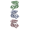
| Deposited unit | 
| ||||||||||||
|---|---|---|---|---|---|---|---|---|---|---|---|---|---|
| 1 |
| ||||||||||||
| Unit cell |
|
-Components
| #1: Protein | Mass: 52963.352 Da / Num. of mol.: 1 Source method: isolated from a genetically manipulated source Source: (gene. exp.) Marinomonas primoryensis (bacteria) / Production host: | ||||||||
|---|---|---|---|---|---|---|---|---|---|
| #2: Protein/peptide | Mass: 436.372 Da / Num. of mol.: 1 / Source method: obtained synthetically / Source: (synth.) synthetic construct (others) | ||||||||
| #3: Chemical | ChemComp-CA /   Mass: 40.078 Da / Num. of mol.: 16 / Source method: obtained synthetically / Formula: Ca Mass: 40.078 Da / Num. of mol.: 16 / Source method: obtained synthetically / Formula: Ca#4: Chemical | ChemComp-1PE / |   Mass: 238.278 Da / Num. of mol.: 1 / Source method: obtained synthetically / Formula: C10H22O6 / Comment: precipitant*YM Mass: 238.278 Da / Num. of mol.: 1 / Source method: obtained synthetically / Formula: C10H22O6 / Comment: precipitant*YM#5: Water | ChemComp-HOH / |  Mass: 18.015 Da / Num. of mol.: 447 / Source method: isolated from a natural source / Formula: H2O Mass: 18.015 Da / Num. of mol.: 447 / Source method: isolated from a natural source / Formula: H2OHas ligand of interest | N | Has protein modification | N | |
-Experimental details
-Experiment
| Experiment | Method: X-RAY DIFFRACTION / Number of used crystals: 1 |
|---|
- Sample preparation
| Crystal | Density Matthews: 1.54 Å3/Da / Density % sol: 20.14 % |
|---|---|
| Crystal grow | Temperature: 298 K / Method: microbatch / pH: 4.6 Details: ~0.1 M calcium chloride, ~0.1 M sodium acetate (pH 4.6), ~30% (w/v) PEG 400 |
-Data collection
| Diffraction | Mean temperature: 93 K / Serial crystal experiment: N |
|---|---|
| Diffraction source | Source: SYNCHROTRON / Site: PETRA III, DESY  / Beamline: P11 / Wavelength: 1.0332 Å / Beamline: P11 / Wavelength: 1.0332 Å |
| Detector | Type: DECTRIS PILATUS 6M / Detector: PIXEL / Date: Apr 7, 2018 |
| Radiation | Protocol: SINGLE WAVELENGTH / Monochromatic (M) / Laue (L): M / Scattering type: x-ray |
| Radiation wavelength | Wavelength: 1.0332 Å / Relative weight: 1 |
| Reflection | Resolution: 1.9→57.42 Å / Num. obs: 48424 / % possible obs: 98 % / Redundancy: 6.9 % / Biso Wilson estimate: 23.98 Å2 / Rmerge(I) obs: 0.068 / Net I/σ(I): 12.8 |
| Reflection shell | Resolution: 2→2.05 Å / Rmerge(I) obs: 1.697 / Num. unique obs: 20688 |
- Processing
| Software |
| |||||||||||||||||||||||||||||||||||||||||||||||||||||||||||||||||||||||||||||||||||||||||||||||||||||||||||||||||||||||||||||||||||||
|---|---|---|---|---|---|---|---|---|---|---|---|---|---|---|---|---|---|---|---|---|---|---|---|---|---|---|---|---|---|---|---|---|---|---|---|---|---|---|---|---|---|---|---|---|---|---|---|---|---|---|---|---|---|---|---|---|---|---|---|---|---|---|---|---|---|---|---|---|---|---|---|---|---|---|---|---|---|---|---|---|---|---|---|---|---|---|---|---|---|---|---|---|---|---|---|---|---|---|---|---|---|---|---|---|---|---|---|---|---|---|---|---|---|---|---|---|---|---|---|---|---|---|---|---|---|---|---|---|---|---|---|---|---|---|
| Refinement | Method to determine structure: MOLECULAR REPLACEMENT Starting model: 58kg Resolution: 1.9→57.39 Å / SU ML: 0.2053 / Cross valid method: FREE R-VALUE / σ(F): 1.98 / Phase error: 24.9194 Stereochemistry target values: GeoStd + Monomer Library + CDL v1.2
| |||||||||||||||||||||||||||||||||||||||||||||||||||||||||||||||||||||||||||||||||||||||||||||||||||||||||||||||||||||||||||||||||||||
| Solvent computation | Shrinkage radii: 0.9 Å / VDW probe radii: 1.11 Å / Solvent model: FLAT BULK SOLVENT MODEL | |||||||||||||||||||||||||||||||||||||||||||||||||||||||||||||||||||||||||||||||||||||||||||||||||||||||||||||||||||||||||||||||||||||
| Displacement parameters | Biso mean: 32.85 Å2 | |||||||||||||||||||||||||||||||||||||||||||||||||||||||||||||||||||||||||||||||||||||||||||||||||||||||||||||||||||||||||||||||||||||
| Refinement step | Cycle: LAST / Resolution: 1.9→57.39 Å
| |||||||||||||||||||||||||||||||||||||||||||||||||||||||||||||||||||||||||||||||||||||||||||||||||||||||||||||||||||||||||||||||||||||
| Refine LS restraints |
| |||||||||||||||||||||||||||||||||||||||||||||||||||||||||||||||||||||||||||||||||||||||||||||||||||||||||||||||||||||||||||||||||||||
| LS refinement shell |
|
