 Movie
Movie Controller

[English] 日本語
 Yorodumi
- PDB-6x45: SARS-CoV2 spike glycoprotein N-terminal heptad repeat domain + SA... -
+ Open data

- Basic information
| Entry | Database: PDB / ID: 6x45 | |||||||||
|---|---|---|---|---|---|---|---|---|---|---|
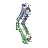
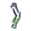
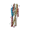
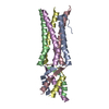
| Title | SARS-CoV2 spike glycoprotein N-terminal heptad repeat domain + SARS-CoV2(QEYKKEKE) | |||||||||
 Components Components | (Spike protein S2') x 2 | |||||||||
 Keywords Keywords | VIRAL PROTEIN / Coronavirus / COVID-19 / SARS-CoV-2 / spike / antiviral | |||||||||
| Function / homology |  Function and homology information Function and homology informationsymbiont-mediated disruption of host tissue / Maturation of spike protein / Translation of Structural Proteins / Virion Assembly and Release / host cell surface / host extracellular region / symbiont-mediated-mediated suppression of host tetherin activity / Induction of Cell-Cell Fusion / structural constituent of virion / positive regulation of viral entry into host cell ...symbiont-mediated disruption of host tissue / Maturation of spike protein / Translation of Structural Proteins / Virion Assembly and Release / host cell surface / host extracellular region / symbiont-mediated-mediated suppression of host tetherin activity / Induction of Cell-Cell Fusion / structural constituent of virion / positive regulation of viral entry into host cell / membrane fusion / host cell endoplasmic reticulum-Golgi intermediate compartment membrane / Attachment and Entry / entry receptor-mediated virion attachment to host cell / receptor-mediated virion attachment to host cell / host cell surface receptor binding / symbiont-mediated suppression of host innate immune response / endocytosis involved in viral entry into host cell / receptor ligand activity / fusion of virus membrane with host plasma membrane / fusion of virus membrane with host endosome membrane / viral envelope / symbiont entry into host cell / virion attachment to host cell / host cell plasma membrane / SARS-CoV-2 activates/modulates innate and adaptive immune responses / virion membrane / membrane / identical protein binding / plasma membrane Similarity search - Function | |||||||||
| Biological species |   Severe acute respiratory syndrome coronavirus 2 Severe acute respiratory syndrome coronavirus 2 | |||||||||
| Method |  X-RAY DIFFRACTION / SYNCHROTRON / MOLECULAR REPLACEMENT / Resolution: 2.2 Å X-RAY DIFFRACTION / SYNCHROTRON / MOLECULAR REPLACEMENT / Resolution: 2.2 Å | |||||||||
 Authors Authors | Kreitler, D.F. / Outlaw, V.K. / Gellman, S.H. | |||||||||
| Funding support |  United States, 2items United States, 2items
| |||||||||
 Citation Citation | Journal: To Be Published Title: Engineered peptides potently block entry of SARS-CoV-2 into human airway cells Authors: Outlaw, V.K. / Yu, Z. / Kreitler, D.F. / Bovier, F. / Porotto, M. / Moscona, A. / Gellman, S.H. | |||||||||
| History |
|
- Structure visualization
| Structure viewer | Molecule: MolmilJmol/JSmol |
|---|
- Downloads & links
-Download
| PDBx/mmCIF format | 6x45.cif.gz | 169.5 KB | Display | PDBx/mmCIF format |
|---|---|---|---|---|
| PDB format | pdb6x45.ent.gz | 127 KB | Display | PDB format |
| PDBx/mmJSON format | 6x45.json.gz | Tree view | PDBx/mmJSON format | |
| Others |  Other downloads Other downloads |
-Validation report
| Arichive directory | https://data.pdbj.org/pub/pdb/validation_reports/x4/6x45ftp://data.pdbj.org/pub/pdb/validation_reports/x4/6x45 | HTTPS FTP |
|---|
-Related structure data
| Related structure data |  6lxtS S: Starting model for refinement |
|---|---|
| Similar structure data | |
| Experimental dataset #1 | Data reference: 10.5281/zenodo.13904737 / Data set type: diffraction image data |


-Links
 PDBj
PDBj


- Assembly
| Deposited unit | 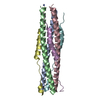
| ||||||||||||
|---|---|---|---|---|---|---|---|---|---|---|---|---|---|
| 1 |
| ||||||||||||
| Unit cell |
|
-Components
| #1: Protein/peptide | Mass: 4172.749 Da / Num. of mol.: 3 Mutation: G1171Q, Q1180E, K1181Y, D1184K, R1185K, N1187E, N1192K, N1194E Source method: obtained synthetically Details: This compound is derived from the SARS-CoV2 spike glycoprotein C-terminal heptad repeat domain residues 1168-1203 with substitutions G1171Q, Q1180E, K1181Y, D1184K, R1185K, N1187E, N1192K, ...Details: This compound is derived from the SARS-CoV2 spike glycoprotein C-terminal heptad repeat domain residues 1168-1203 with substitutions G1171Q, Q1180E, K1181Y, D1184K, R1185K, N1187E, N1192K, N1194E. It is acetylated at the N-terminus and amidated at the C-terminus. Source: (synth.) Severe acute respiratory syndrome coronavirus 2References: UniProt: P0DTC2 #2: Protein | Mass: 5972.696 Da / Num. of mol.: 3 / Source method: obtained synthetically Details: This compound is derived from the SARS-CoV2 spike glycoprotein N-terminal heptad repeat domain residues 912-966. It is acetylated at the N-terminus and amidated at the C-terminus. Source: (synth.) Severe acute respiratory syndrome coronavirus 2References: UniProt: P0DTC2 #3: Water | ChemComp-HOH / |  Mass: 18.015 Da / Num. of mol.: 8 / Source method: isolated from a natural source / Formula: H2O Mass: 18.015 Da / Num. of mol.: 8 / Source method: isolated from a natural source / Formula: H2OHas ligand of interest | N | Has protein modification | Y | |
|---|
-Experimental details
-Experiment
| Experiment | Method: X-RAY DIFFRACTION / Number of used crystals: 1 |
|---|
- Sample preparation
| Crystal | Density Matthews: 1.92 Å3/Da / Density % sol: 35.9 % / Description: 3D bricks |
|---|---|
| Crystal grow | Temperature: 293 K / Method: vapor diffusion, hanging drop / pH: 7.8 Details: Peptide solution: Lyophilized TFA salt dissolved in 1% w/v beta-D-octylglucoside; HRN (4 mg/mL), HRC-QEYKKEKE (3 mg/mL) Well solution: 0.1 M Tris pH 7.8, 26% w/v PEG3350, 0.3 M MgCl2 Drop: 1 ...Details: Peptide solution: Lyophilized TFA salt dissolved in 1% w/v beta-D-octylglucoside; HRN (4 mg/mL), HRC-QEYKKEKE (3 mg/mL) Well solution: 0.1 M Tris pH 7.8, 26% w/v PEG3350, 0.3 M MgCl2 Drop: 1 uL peptide solution, 1 uL well solution Reservoir: 150 uL in VDXm plate |
-Data collection
| Diffraction | Mean temperature: 100 K / Serial crystal experiment: N |
|---|---|
| Diffraction source | Source: SYNCHROTRON / Site: NSLS-II / Beamline: 17-ID-1 / Wavelength: 0.920087 Å |
| Detector | Type: DECTRIS EIGER X 9M / Detector: PIXEL / Date: May 6, 2020 |
| Radiation | Monochromator: Si(111) double crystal / Protocol: SINGLE WAVELENGTH / Monochromatic (M) / Laue (L): M / Scattering type: x-ray |
| Radiation wavelength | Wavelength: 0.920087 Å / Relative weight: 1 |
| Reflection | Resolution: 2.2→80.12 Å / Num. obs: 12351 / % possible obs: 99.5 % / Redundancy: 6.5 % / Biso Wilson estimate: 54.79 Å2 / CC1/2: 0.999 / Rpim(I) all: 0.038 / Rrim(I) all: 0.096 / Rsym value: 0.088 / Net I/σ(I): 9 |
| Reflection shell | Resolution: 2.2→2.27 Å / Redundancy: 6.3 % / Mean I/σ(I) obs: 1.2 / Num. unique obs: 1047 / CC1/2: 0.475 / Rpim(I) all: 0.666 / Rrim(I) all: 1.705 / Rsym value: 1.565 / % possible all: 99.2 |
- Processing
| Software |
| ||||||||||||||||||||||||||||||||||||||||||||||||||||||||||||||||||||||
|---|---|---|---|---|---|---|---|---|---|---|---|---|---|---|---|---|---|---|---|---|---|---|---|---|---|---|---|---|---|---|---|---|---|---|---|---|---|---|---|---|---|---|---|---|---|---|---|---|---|---|---|---|---|---|---|---|---|---|---|---|---|---|---|---|---|---|---|---|---|---|---|
| Refinement | Method to determine structure: MOLECULAR REPLACEMENT Starting model: 6LXT Resolution: 2.2→45.04 Å / SU ML: 0.327 / Cross valid method: FREE R-VALUE / σ(F): 1.34 / Phase error: 30.8919 Stereochemistry target values: GeoStd + Monomer Library + CDL v1.2
| ||||||||||||||||||||||||||||||||||||||||||||||||||||||||||||||||||||||
| Solvent computation | Shrinkage radii: 0.9 Å / VDW probe radii: 1.11 Å / Solvent model: FLAT BULK SOLVENT MODEL | ||||||||||||||||||||||||||||||||||||||||||||||||||||||||||||||||||||||
| Displacement parameters | Biso mean: 88.63 Å2 | ||||||||||||||||||||||||||||||||||||||||||||||||||||||||||||||||||||||
| Refinement step | Cycle: LAST / Resolution: 2.2→45.04 Å
| ||||||||||||||||||||||||||||||||||||||||||||||||||||||||||||||||||||||
| Refine LS restraints |
| ||||||||||||||||||||||||||||||||||||||||||||||||||||||||||||||||||||||
| LS refinement shell |
| ||||||||||||||||||||||||||||||||||||||||||||||||||||||||||||||||||||||
| Refinement TLS params. | Method: refined / Origin x: 14.4060869838 Å / Origin y: 5.33483149575 Å / Origin z: 37.9270700186 Å
| ||||||||||||||||||||||||||||||||||||||||||||||||||||||||||||||||||||||
| Refinement TLS group | Selection details: all |
