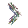
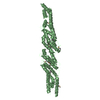
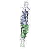
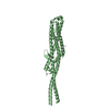
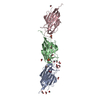
Spikeglycoprotein / S glycoprotein / E2 / Peplomer protein
Mass: 3869.315 Da / Num. of mol.: 3 / Fragment: RESIDUES 914-949 / Source method: obtained synthetically Details: N-TERMINAL CAPPED WITH ACETYL GROUP BUT ONLY VISIBLE DENSITY ON B, C, D AND F. C-TERMINAL A, B, C, D, E, F CAPPED WITH AMINE GROUP Source: (synth.) Human SARS coronavirus / References: UniProt: P59594
#2: Protein/peptide
Spikeglycoprotein / S glycoprotein / E2 / Peplomer protein
Mass: 5217.883 Da / Num. of mol.: 3 / Fragment: RESIDUES 1148-1193 / Source method: obtained synthetically Details: N-TERMINAL CAPPED WITH ACETYL GROUP BUT ONLY VISIBLE DENSITY ON B, C, D AND F. C-TERMINAL A, B, C, D, E, F CAPPED WITH AMINE GROUP Source: (synth.) Human SARS coronavirus / References: UniProt: P59594
Protocol: SINGLE WAVELENGTH / Monochromatic (M) / Laue (L): M / Scattering type: x-ray
Radiation wavelength
Wavelength: 0.934 Å / Relative weight: 1
Reflection
Resolution: 1.6→30 Å / Num. obs: 28450 / % possible obs: 99.6 % / Redundancy: 3.5 % / Rmerge(I) obs: 0.04 / Net I/σ(I): 15
Reflection shell
Resolution: 1.6→1.66 Å / Redundancy: 3.1 % / Rmerge(I) obs: 0.4 / Mean I/σ(I) obs: 3 / % possible all: 93.5
-
Processing
Software
Name
Version
Classification
DENZO
datareduction
SCALEPACK
datascaling
CNS
phasing
ARP/wARP
phasing
REFMAC
5.1.24
refinement
Refinement
Method to determine structure: SIRAS / Resolution: 1.6→30 Å / Cor.coef. Fo:Fc: 0.966 / Cor.coef. Fo:Fc free: 0.952 / SU B: 2.43 / SU ML: 0.081 / Cross valid method: THROUGHOUT / ESU R: 0.105 / ESU R Free: 0.107 / Stereochemistry target values: MAXIMUM LIKELIHOOD Details: HYDROGENS HAVE BEEN ADDED IN THE RIDING POSITIONS. NO ELECTRON DENSITY WAS PRESENT FOR LEU1148 SIDE CHAIN ON CHAIN E AND FOR THE N-TERMINAL CAPPING ACETYL GROUPS OF CHAINS A AND E. THE ...Details: HYDROGENS HAVE BEEN ADDED IN THE RIDING POSITIONS. NO ELECTRON DENSITY WAS PRESENT FOR LEU1148 SIDE CHAIN ON CHAIN E AND FOR THE N-TERMINAL CAPPING ACETYL GROUPS OF CHAINS A AND E. THE CORRESPONDING ATOMS WERE NOT INCLUDED IN THE FINAL MODEL.
Rfactor
Num. reflection
% reflection
Selection details
Rfree
0.241
1521
5.1 %
RANDOM
Rwork
0.199
-
-
-
obs
0.201
28450
99.7 %
-
Solvent computation
Ion probe radii: 0.8 Å / Shrinkage radii: 0.8 Å / VDW probe radii: 1.4 Å / Solvent model: BABINET MODEL WITH MASK
 Movie
Movie Controller

 Yorodumi
 Open data
 Basic information
 Components
 Keywords
 Function and homology information
 Human SARS coronavirus
 X-RAY DIFFRACTION /
  Authors
 Citation
 Structure visualization
 Downloads & links
 Other downloads
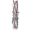


 PDBj
PDBj

 Assembly
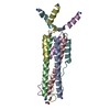

 Mass: 18.015 Da / Num. of mol.: 202 / Source method: isolated from a natural source / Formula: H2O
 Sample preparation
 / Beamline: ID14-1 / Wavelength: 0.934
  Processing