 Movie
Movie Controller

[English] 日本語
 Yorodumi
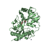
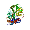
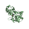
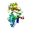
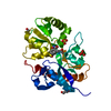
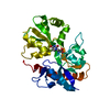
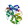
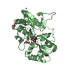
- PDB-6x2d: Crystal Structure of DNase I Domain of Ribonuclease E from Vibrio... -
+ Open data

- Basic information
| Entry | Database: PDB / ID: 6x2d | ||||||
|---|---|---|---|---|---|---|---|
| Title | Crystal Structure of DNase I Domain of Ribonuclease E from Vibrio cholerae | ||||||
 Components Components | Ribonuclease E | ||||||
 Keywords Keywords | RNA BINDING PROTEIN / Structural Genomics / Center for Structural Genomics of Infectious Diseases / CSGID | ||||||
| Function / homology |  Function and homology information Function and homology informationribonuclease E / ribonuclease E activity / tRNA processing / mRNA catabolic process / RNA nuclease activity / RNA endonuclease activity / cytoplasmic side of plasma membrane / rRNA processing / tRNA binding / rRNA binding ...ribonuclease E / ribonuclease E activity / tRNA processing / mRNA catabolic process / RNA nuclease activity / RNA endonuclease activity / cytoplasmic side of plasma membrane / rRNA processing / tRNA binding / rRNA binding / magnesium ion binding / zinc ion binding / cytoplasm Similarity search - Function | ||||||
| Biological species |  Vibrio cholerae serotype O1 (bacteria) Vibrio cholerae serotype O1 (bacteria) | ||||||
| Method |  X-RAY DIFFRACTION / SYNCHROTRON / MOLECULAR REPLACEMENT / Resolution: 1.85 Å X-RAY DIFFRACTION / SYNCHROTRON / MOLECULAR REPLACEMENT / Resolution: 1.85 Å | ||||||
 Authors Authors | Minasov, G. / Shuvalova, L. / Kiryukhina, O. / Dubrovska, I. / Wiersum, G. / Satchell, K.J.F. / Center for Structural Genomics of Infectious Diseases (CSGID) | ||||||
 Citation Citation | Journal: To Be Published Title: Crystal Structure of DNase I Domain of Ribonuclease E from Vibrio cholerae. Authors: Minasov, G. / Shuvalova, L. / Kiryukhina, O. / Dubrovska, I. / Wiersum, G. / Satchell, K.J.F. / Center for Structural Genomics of Infectious Diseases (CSGID) | ||||||
| History |
|
- Structure visualization
| Structure viewer | Molecule: MolmilJmol/JSmol |
|---|
- Downloads & links
-Download
| PDBx/mmCIF format | 6x2d.cif.gz | 101.8 KB | Display | PDBx/mmCIF format |
|---|---|---|---|---|
| PDB format | pdb6x2d.ent.gz | 78.4 KB | Display | PDB format |
| PDBx/mmJSON format | 6x2d.json.gz | Tree view | PDBx/mmJSON format | |
| Others |  Other downloads Other downloads |
-Validation report
| Arichive directory | https://data.pdbj.org/pub/pdb/validation_reports/x2/6x2dftp://data.pdbj.org/pub/pdb/validation_reports/x2/6x2d | HTTPS FTP |
|---|
-Related structure data
| Related structure data |  2vrtS S: Starting model for refinement |
|---|---|
| Similar structure data | |
| Other databases |

-Links
 PDBj
PDBj


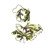
- Assembly
| Deposited unit | 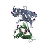
| ||||||||
|---|---|---|---|---|---|---|---|---|---|
| 1 |
| ||||||||
| Unit cell |
|
-Components
| #1: Protein | Mass: 12974.358 Da / Num. of mol.: 2 Source method: isolated from a genetically manipulated source Source: (gene. exp.) Vibrio cholerae serotype O1 (strain ATCC 39315 / El Tor Inaba N16961) (bacteria)Strain: ATCC 39315 / El Tor Inaba N16961 / Gene: rne, VC_2030 / Plasmid: pMCSG53 / Production host: #2: Chemical | ChemComp-IOD /   Mass: 126.904 Da / Num. of mol.: 7 / Source method: obtained synthetically / Formula: I Mass: 126.904 Da / Num. of mol.: 7 / Source method: obtained synthetically / Formula: I#3: Water | ChemComp-HOH / |  Mass: 18.015 Da / Num. of mol.: 73 / Source method: isolated from a natural source / Formula: H2O Mass: 18.015 Da / Num. of mol.: 73 / Source method: isolated from a natural source / Formula: H2OHas ligand of interest | N | Has protein modification | Y | |
|---|
-Experimental details
-Experiment
| Experiment | Method: X-RAY DIFFRACTION / Number of used crystals: 1 |
|---|
- Sample preparation
| Crystal | Density Matthews: 2.06 Å3/Da / Density % sol: 40.4 % |
|---|---|
| Crystal grow | Temperature: 292 K / Method: vapor diffusion, sitting drop / pH: 6.5 Details: Protein: 7.9 mg/ml, 0.01M Tris pH 8.3; Screen: PACT (F3), 0.2M Sodium iodide, 0.1M Bis-Tris propane pH 6.5, 20% (w/v) PEG 3350 |
-Data collection
| Diffraction | Mean temperature: 100 K / Serial crystal experiment: N |
|---|---|
| Diffraction source | Source: SYNCHROTRON / Site: APS  / Beamline: 21-ID-F / Wavelength: 0.97872 Å / Beamline: 21-ID-F / Wavelength: 0.97872 Å |
| Detector | Type: MARMOSAIC 300 mm CCD / Detector: CCD / Date: Mar 17, 2020 / Details: Be |
| Radiation | Monochromator: DIAMOND (111) / Protocol: SINGLE WAVELENGTH / Monochromatic (M) / Laue (L): M / Scattering type: x-ray |
| Radiation wavelength | Wavelength: 0.97872 Å / Relative weight: 1 |
| Reflection | Resolution: 1.85→30 Å / Num. obs: 17863 / % possible obs: 99.4 % / Observed criterion σ(I): -3 / Redundancy: 5.7 % / Biso Wilson estimate: 34.1 Å2 / CC1/2: 0.985 / CC star: 0.996 / Rmerge(I) obs: 0.088 / Rpim(I) all: 0.041 / Rrim(I) all: 0.097 / Rsym value: 0.088 / Χ2: 1.466 / Net I/σ(I): 21.9 |
| Reflection shell | Resolution: 1.85→1.88 Å / Redundancy: 6 % / Rmerge(I) obs: 0.759 / Mean I/σ(I) obs: 4.7 / Num. unique obs: 897 / CC1/2: 0.927 / CC star: 0.981 / Rpim(I) all: 0.339 / Rrim(I) all: 0.833 / Rsym value: 0.759 / % possible all: 100 |
- Processing
| Software |
| |||||||||||||||||||||||||||||||||||||||||||||||||||||||||||||||||||||||||||||||||||||||||||||||||||||||||||||||||||||||||||||||||||||||||||||||||||||||||||||||||||||||||||||||
|---|---|---|---|---|---|---|---|---|---|---|---|---|---|---|---|---|---|---|---|---|---|---|---|---|---|---|---|---|---|---|---|---|---|---|---|---|---|---|---|---|---|---|---|---|---|---|---|---|---|---|---|---|---|---|---|---|---|---|---|---|---|---|---|---|---|---|---|---|---|---|---|---|---|---|---|---|---|---|---|---|---|---|---|---|---|---|---|---|---|---|---|---|---|---|---|---|---|---|---|---|---|---|---|---|---|---|---|---|---|---|---|---|---|---|---|---|---|---|---|---|---|---|---|---|---|---|---|---|---|---|---|---|---|---|---|---|---|---|---|---|---|---|---|---|---|---|---|---|---|---|---|---|---|---|---|---|---|---|---|---|---|---|---|---|---|---|---|---|---|---|---|---|---|---|---|---|
| Refinement | Method to determine structure: MOLECULAR REPLACEMENT Starting model: 2vrt Resolution: 1.85→29.41 Å / Cor.coef. Fo:Fc: 0.956 / Cor.coef. Fo:Fc free: 0.942 / SU B: 9.97 / SU ML: 0.142 / Cross valid method: THROUGHOUT / σ(F): 0 / ESU R: 0.169 / ESU R Free: 0.15 / Stereochemistry target values: MAXIMUM LIKELIHOOD Details: HYDROGENS HAVE BEEN ADDED IN THE RIDING POSITIONS U VALUES : WITH TLS ADDED
| |||||||||||||||||||||||||||||||||||||||||||||||||||||||||||||||||||||||||||||||||||||||||||||||||||||||||||||||||||||||||||||||||||||||||||||||||||||||||||||||||||||||||||||||
| Solvent computation | Ion probe radii: 0.8 Å / Shrinkage radii: 0.8 Å / VDW probe radii: 1.2 Å / Solvent model: MASK | |||||||||||||||||||||||||||||||||||||||||||||||||||||||||||||||||||||||||||||||||||||||||||||||||||||||||||||||||||||||||||||||||||||||||||||||||||||||||||||||||||||||||||||||
| Displacement parameters | Biso max: 103.88 Å2 / Biso mean: 43.735 Å2 / Biso min: 27.73 Å2
| |||||||||||||||||||||||||||||||||||||||||||||||||||||||||||||||||||||||||||||||||||||||||||||||||||||||||||||||||||||||||||||||||||||||||||||||||||||||||||||||||||||||||||||||
| Refinement step | Cycle: final / Resolution: 1.85→29.41 Å
| |||||||||||||||||||||||||||||||||||||||||||||||||||||||||||||||||||||||||||||||||||||||||||||||||||||||||||||||||||||||||||||||||||||||||||||||||||||||||||||||||||||||||||||||
| Refine LS restraints |
| |||||||||||||||||||||||||||||||||||||||||||||||||||||||||||||||||||||||||||||||||||||||||||||||||||||||||||||||||||||||||||||||||||||||||||||||||||||||||||||||||||||||||||||||
| LS refinement shell | Resolution: 1.852→1.9 Å / Rfactor Rfree error: 0 / Total num. of bins used: 20
| |||||||||||||||||||||||||||||||||||||||||||||||||||||||||||||||||||||||||||||||||||||||||||||||||||||||||||||||||||||||||||||||||||||||||||||||||||||||||||||||||||||||||||||||
| Refinement TLS params. | Method: refined / Refine-ID: X-RAY DIFFRACTION
| |||||||||||||||||||||||||||||||||||||||||||||||||||||||||||||||||||||||||||||||||||||||||||||||||||||||||||||||||||||||||||||||||||||||||||||||||||||||||||||||||||||||||||||||
| Refinement TLS group |
|
