 Movie
Movie Controller

[English] 日本語
 Yorodumi
- PDB-6x1r: Crystal Structure of Choanoflagellate (Monosiga brevicollis) Dlg1... -
+ Open data

- Basic information
| Entry | Database: PDB / ID: 6x1r | ||||||
|---|---|---|---|---|---|---|---|
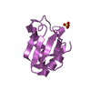
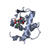
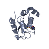
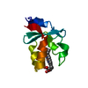
| Title | Crystal Structure of Choanoflagellate (Monosiga brevicollis) Dlg1 PDZ2 (mbDLG-2) in spacegroup P212121 | ||||||
 Components Components | mbDLG protein | ||||||
 Keywords Keywords | SIGNALING PROTEIN / PDZ / protein-protein interaction / choanoflagellates / Monosiga brevicollis / peptide-binding domain | ||||||
| Function / homology |  Function and homology information Function and homology informationreceptor localization to synapse / establishment or maintenance of epithelial cell apical/basal polarity / receptor clustering / cell-cell adhesion / cell junction / basolateral plasma membrane / protein kinase binding Similarity search - Function | ||||||
| Biological species |  | ||||||
| Method |  X-RAY DIFFRACTION / SYNCHROTRON / MOLECULAR REPLACEMENT / Resolution: 1.3 Å X-RAY DIFFRACTION / SYNCHROTRON / MOLECULAR REPLACEMENT / Resolution: 1.3 Å | ||||||
 Authors Authors | Bamonte, H.A. / Amacher, J.F. | ||||||
| Funding support |  United States, 1items United States, 1items
| ||||||
 Citation Citation | Journal: Protein Sci. / Year: 2020 Title: Structural characterization and computational analysis of PDZ domains in Monosiga brevicollis. Authors: Gao, M. / Mackley, I.G.P. / Mesbahi-Vasey, S. / Bamonte, H.A. / Struyvenberg, S.A. / Landolt, L. / Pederson, N.J. / Williams, L.I. / Bahl, C.D. / Brooks 3rd, L. / Amacher, J.F. | ||||||
| History |
|
- Structure visualization
| Structure viewer | Molecule: MolmilJmol/JSmol |
|---|
- Downloads & links
-Download
| PDBx/mmCIF format | 6x1r.cif.gz | 35.6 KB | Display | PDBx/mmCIF format |
|---|---|---|---|---|
| PDB format | pdb6x1r.ent.gz | 22.2 KB | Display | PDB format |
| PDBx/mmJSON format | 6x1r.json.gz | Tree view | PDBx/mmJSON format | |
| Others |  Other downloads Other downloads |
-Validation report
| Arichive directory | https://data.pdbj.org/pub/pdb/validation_reports/x1/6x1rftp://data.pdbj.org/pub/pdb/validation_reports/x1/6x1r | HTTPS FTP |
|---|
-Related structure data
| Related structure data | 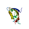 6x1nC  6x1pC 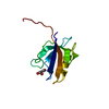 6x1xC 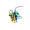 6x20C 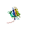 6x22C  6x23C  2bygS S: Starting model for refinement C: citing same article ( |
|---|---|
| Similar structure data |

-Links
 PDBj
PDBj- Assembly
| Deposited unit | 
| ||||||||
|---|---|---|---|---|---|---|---|---|---|
| 1 |
| ||||||||
| Unit cell |
|
-Components
| #1: Protein | Mass: 10847.223 Da / Num. of mol.: 1 / Fragment: second PDZ domain (UNP residues 93-192) Source method: isolated from a genetically manipulated source Source: (gene. exp.)  |
|---|---|
| #2: Chemical | ChemComp-GOL /   Mass: 92.094 Da / Num. of mol.: 1 / Source method: obtained synthetically / Formula: C3H8O3 Mass: 92.094 Da / Num. of mol.: 1 / Source method: obtained synthetically / Formula: C3H8O3 |
| #3: Water | ChemComp-HOH /  Mass: 18.015 Da / Num. of mol.: 123 / Source method: isolated from a natural source / Formula: H2O Mass: 18.015 Da / Num. of mol.: 123 / Source method: isolated from a natural source / Formula: H2O |
| Has ligand of interest | N |
-Experimental details
-Experiment
| Experiment | Method: X-RAY DIFFRACTION / Number of used crystals: 1 |
|---|
- Sample preparation
| Crystal | Density Matthews: 2.63 Å3/Da / Density % sol: 53.28 % |
|---|---|
| Crystal grow | Temperature: 298 K / Method: vapor diffusion, hanging drop / pH: 5 / Details: 0.2 M sodium malonate, pH 5, 20% w/v PEG3350 |
-Data collection
| Diffraction | Mean temperature: 80 K / Serial crystal experiment: N | ||||||||||||||||||
|---|---|---|---|---|---|---|---|---|---|---|---|---|---|---|---|---|---|---|---|
| Diffraction source | Source: SYNCHROTRON / Site: ALS / Beamline: 5.0.1 / Wavelength: 0.97741 Å | ||||||||||||||||||
| Detector | Type: DECTRIS PILATUS3 6M / Detector: PIXEL / Date: Mar 12, 2020 | ||||||||||||||||||
| Radiation | Monochromator: Si(220) / Protocol: SINGLE WAVELENGTH / Monochromatic (M) / Laue (L): M / Scattering type: x-ray | ||||||||||||||||||
| Radiation wavelength | Wavelength: 0.97741 Å / Relative weight: 1 | ||||||||||||||||||
| Reflection | Resolution: 1.3→40.3 Å / Num. obs: 28809 / % possible obs: 99.4 % / Redundancy: 6.04 % / Biso Wilson estimate: 24.3 Å2 / CC1/2: 1 / Rsym value: 0.033 / Net I/σ(I): 24.04 | ||||||||||||||||||
| Reflection shell | CC1/2: 0.866 / Diffraction-ID: 1
|
- Processing
| Software |
| ||||||||||||||||||||||||||||||||||||||||||||||||||||||||||||||||||
|---|---|---|---|---|---|---|---|---|---|---|---|---|---|---|---|---|---|---|---|---|---|---|---|---|---|---|---|---|---|---|---|---|---|---|---|---|---|---|---|---|---|---|---|---|---|---|---|---|---|---|---|---|---|---|---|---|---|---|---|---|---|---|---|---|---|---|---|
| Refinement | Method to determine structure: MOLECULAR REPLACEMENT Starting model: PDB entry 2BYG Resolution: 1.3→40.299 Å / SU ML: 0.13 / Cross valid method: THROUGHOUT / σ(F): 1.35 / Phase error: 21.66 / Stereochemistry target values: ML
| ||||||||||||||||||||||||||||||||||||||||||||||||||||||||||||||||||
| Solvent computation | Shrinkage radii: 0.9 Å / VDW probe radii: 1.11 Å / Solvent model: FLAT BULK SOLVENT MODEL | ||||||||||||||||||||||||||||||||||||||||||||||||||||||||||||||||||
| Displacement parameters | Biso max: 75.48 Å2 / Biso mean: 24.4252 Å2 / Biso min: 11.85 Å2 | ||||||||||||||||||||||||||||||||||||||||||||||||||||||||||||||||||
| Refinement step | Cycle: final / Resolution: 1.3→40.299 Å
| ||||||||||||||||||||||||||||||||||||||||||||||||||||||||||||||||||
| Refine LS restraints |
| ||||||||||||||||||||||||||||||||||||||||||||||||||||||||||||||||||
| LS refinement shell | Refine-ID: X-RAY DIFFRACTION / Rfactor Rfree error: 0
|
