 Movie
Movie Controller

[English] 日本語
 Yorodumi
- PDB-6wmc: Crystal structure of a soluble variant of full-length human APOBE... -
+ Open data

- Basic information
| Entry | Database: PDB / ID: 6wmc | ||||||
|---|---|---|---|---|---|---|---|
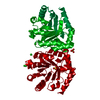
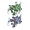
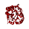
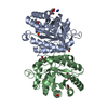
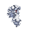
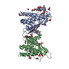
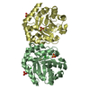
| Title | Crystal structure of a soluble variant of full-length human APOBEC3G (pH 9.0) | ||||||
 Components Components |
| ||||||
 Keywords Keywords | HYDROLASE/DNA / APOBEC3G / ANTIVIRAL DEFENSE / HYDROLASE / DNA CYTIDINE DEAMINASE / HYDROLASE-DNA complex | ||||||
| Function / homology | Cytidine Deaminase, domain 2 / Cytidine Deaminase; domain 2 / 3-Layer(aba) Sandwich / Alpha Beta / DNA Function and homology information Function and homology information | ||||||
| Biological species |  Homo sapiens (human) Homo sapiens (human) | ||||||
| Method |  X-RAY DIFFRACTION / SYNCHROTRON / MOLECULAR REPLACEMENT / Resolution: 3.49 Å X-RAY DIFFRACTION / SYNCHROTRON / MOLECULAR REPLACEMENT / Resolution: 3.49 Å | ||||||
 Authors Authors | Maiti, A. / Matsuo, H. | ||||||
| Funding support |  United States, 1items United States, 1items
| ||||||
 Citation Citation | Journal: J.Mol.Biol. / Year: 2020 Title: Crystal Structure of a Soluble APOBEC3G Variant Suggests ssDNA to Bind in a Channel that Extends between the Two Domains. Authors: Maiti, A. / Myint, W. / Delviks-Frankenberry, K.A. / Hou, S. / Kanai, T. / Balachandran, V. / Sierra Rodriguez, C. / Tripathi, R. / Kurt Yilmaz, N. / Pathak, V.K. / Schiffer, C.A. / Matsuo, H. | ||||||
| History |
| ||||||
| Remark 650 | HELIX DETERMINATION METHOD: AUTHOR | ||||||
| Remark 700 | SHEET DETERMINATION METHOD: AUTHOR |
- Structure visualization
| Structure viewer | Molecule: MolmilJmol/JSmol |
|---|
- Downloads & links
-Download
| PDBx/mmCIF format | 6wmc.cif.gz | 103.6 KB | Display | PDBx/mmCIF format |
|---|---|---|---|---|
| PDB format | pdb6wmc.ent.gz | 65.1 KB | Display | PDB format |
| PDBx/mmJSON format | 6wmc.json.gz | Tree view | PDBx/mmJSON format | |
| Others |  Other downloads Other downloads |
-Validation report
| Arichive directory | https://data.pdbj.org/pub/pdb/validation_reports/wm/6wmcftp://data.pdbj.org/pub/pdb/validation_reports/wm/6wmc | HTTPS FTP |
|---|
-Related structure data
| Related structure data | 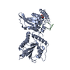 6wmaC 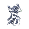 6wmbC 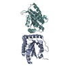 5k81S 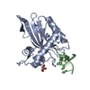 6buxS S: Starting model for refinement C: citing same article ( |
|---|---|
| Similar structure data |


-Links
 PDBj
PDBj- Assembly
| Deposited unit | 
| ||||||||||||
|---|---|---|---|---|---|---|---|---|---|---|---|---|---|
| 1 |
| ||||||||||||
| Unit cell |
|
-Components
| #1: Protein | Mass: 42467.715 Da / Num. of mol.: 1 Source method: isolated from a genetically manipulated source Source: (gene. exp.) Homo sapiens (human) / Gene: APOBEC3G / Production host: |
|---|---|
| #2: DNA chain | Mass: 533.406 Da / Num. of mol.: 1 Source method: isolated from a genetically manipulated source Source: (gene. exp.) |
| #3: Chemical | ChemComp-ZN /   Mass: 65.409 Da / Num. of mol.: 1 / Source method: obtained synthetically / Formula: Zn Mass: 65.409 Da / Num. of mol.: 1 / Source method: obtained synthetically / Formula: Zn |
| #4: Water | ChemComp-HOH /  Mass: 18.015 Da / Num. of mol.: 39 / Source method: isolated from a natural source / Formula: H2O Mass: 18.015 Da / Num. of mol.: 39 / Source method: isolated from a natural source / Formula: H2O |
| Has ligand of interest | N |
-Experimental details
-Experiment
| Experiment | Method: X-RAY DIFFRACTION / Number of used crystals: 1 |
|---|
- Sample preparation
| Crystal | Density Matthews: 2.88 Å3/Da / Density % sol: 57.33 % |
|---|---|
| Crystal grow | Temperature: 277 K / Method: vapor diffusion, sitting drop / pH: 9 / Details: 0.1M BICINE (pH 9.0) and 10% w/v PEG 6000 |
-Data collection
| Diffraction | Mean temperature: 100 K / Serial crystal experiment: N |
|---|---|
| Diffraction source | Source: SYNCHROTRON / Site: APS / Beamline: 22-BM / Wavelength: 1 Å |
| Detector | Type: RAYONIX MX300-HS / Detector: CCD / Date: Feb 8, 2019 |
| Radiation | Protocol: SINGLE WAVELENGTH / Monochromatic (M) / Laue (L): M / Scattering type: x-ray |
| Radiation wavelength | Wavelength: 1 Å / Relative weight: 1 |
| Reflection | Resolution: 3.49→50 Å / Num. obs: 5588 / % possible obs: 89 % / Redundancy: 2.8 % / Biso Wilson estimate: 69.24 Å2 / CC1/2: 0.985 / Rmerge(I) obs: 0.158 / Net I/σ(I): 5.08 |
| Reflection shell | Resolution: 3.49→3.63 Å / Redundancy: 2.6 % / Rmerge(I) obs: 0.497 / Mean I/σ(I) obs: 1.37 / Num. unique obs: 482 / CC1/2: 0.718 / % possible all: 83.4 |
- Processing
| Software |
| |||||||||||||||||||||||||||||||||||
|---|---|---|---|---|---|---|---|---|---|---|---|---|---|---|---|---|---|---|---|---|---|---|---|---|---|---|---|---|---|---|---|---|---|---|---|---|
| Refinement | Method to determine structure: MOLECULAR REPLACEMENT Starting model: 6BUX CHAIN A AND 5K81 CHAIN A Resolution: 3.49→47.72 Å / SU ML: 0.5317 / Cross valid method: FREE R-VALUE / σ(F): 1.33 / Phase error: 28.896
| |||||||||||||||||||||||||||||||||||
| Solvent computation | Shrinkage radii: 0.9 Å / VDW probe radii: 1.11 Å | |||||||||||||||||||||||||||||||||||
| Displacement parameters | Biso mean: 66.28 Å2 | |||||||||||||||||||||||||||||||||||
| Refinement step | Cycle: LAST / Resolution: 3.49→47.72 Å
| |||||||||||||||||||||||||||||||||||
| Refine LS restraints |
| |||||||||||||||||||||||||||||||||||
| LS refinement shell |
|
