Mass: 18.015 Da / Num. of mol.: 229 / Source method: isolated from a natural source / Formula: H2O
-
Details
Has ligand of interest
Y
-
Experimental details
-
Experiment
Experiment
Method: X-RAY DIFFRACTION / Number of used crystals: 3
-
Sample preparation
Crystal
ID
Density Matthews (Å3/Da)
Density % sol (%)
1
2.85
56.85
2
3
Crystal grow
Temperature (K)
Crystal-ID
Method
pH
Details
277
1
vapor diffusion, sitting drop
7.5
0.2 M magnesium chloride, 30% PEG 400, 0.1 M HEPES, pH 7.5. Additive: 0.002 M manganese chloride, 0.01 M UDP-Galactose
277
2
vapor diffusion, sitting drop
7.5
0.2 M magnesium chloride, 30% PEG 400, 0.1 M HEPES, pH 7.5. Additive: 0.002 M manganese chloride, 0.01 M UDP-Galactose, crystal soaked in higher Mn and UDP-Glc prior to flash cooling.
277
3
vapor diffusion, sitting drop
7.5
0.2 M magnesium chloride, 30% PEG 400, 0.1 M HEPES, pH 7.5. Additive: 0.002 M manganese chloride, 0.01 M UDP-Galactose. Crystal soaked in 1 M NaI prior to flash cooling
-
Data collection
Diffraction
ID
Mean temperature (K)
Crystal-ID
Serial crystal experiment
1
100
1
N
2
100
2
N
3
100
3
N
Diffraction source
Source
Site
Beamline
ID
Wavelength (Å)
SYNCHROTRON
ALS
8.3.1
1
1.1271
SYNCHROTRON
ALS
8.3.1
2
1.1158
SYNCHROTRON
ALS
8.3.1
3
1.7136
Detector
Type
ID
Detector
Date
DECTRIS PILATUS3 6M
1
PIXEL
Jun 2, 2016
DECTRIS PILATUS3 6M
2
PIXEL
Oct 12, 2016
DECTRIS PILATUS3 6M
3
PIXEL
Sep 15, 2016
Radiation
ID
Monochromator
Protocol
Monochromatic (M) / Laue (L)
Scattering type
Wavelength-ID
1
Si(111)
SINGLEWAVELENGTH
M
x-ray
1
2
Si(111)
SINGLEWAVELENGTH
M
x-ray
2
3
Si(111)
SINGLEWAVELENGTH
M
x-ray
3
Radiation wavelength
ID
Wavelength (Å)
Relative weight
1
1.1271
1
2
1.1158
1
3
1.7136
1
Reflection
Biso Wilson estimate: 31.679 Å2 / Entry-ID: 6WFV
Resolution (Å)
Num. obs
% possible obs (%)
Redundancy (%)
CC1/2
Rmerge(I) obs
Rrim(I) all
Χ2
Diffraction-ID
Net I/σ(I)
1.7-41.18
36393
99.7
17.521
0.999
0.117
0.12
0.886
1
19.67
1.96-41.2
22942
96.4
16.4
1
0.037
0.038
2
55.2
2.23-61.5
13578
83.7
29.6
1
0.131
0.134
3
22.8
Reflection shell
Diffraction-ID: 1
Resolution (Å)
Redundancy (%)
Rmerge(I) obs
Mean I/σ(I) obs
Num. measured obs
Num. possible
Num. unique obs
CC1/2
Rrim(I) all
% possible all
1.7-1.74
9.969
1.64
2.1
25451
2649
2553
0.717
1.73
96.4
1.74-1.79
12.41
1.349
3.04
32216
2600
2596
0.869
1.407
99.8
1.79-1.84
16.304
1.088
4.57
41234
2529
2529
0.947
1.123
100
1.84-1.9
19.388
0.868
6.4
47171
2434
2433
0.977
0.891
100
1.9-1.96
19.111
0.681
8.19
45867
2400
2400
0.985
0.7
100
1.96-2.03
18.821
0.495
10.35
43251
2298
2298
0.99
0.508
100
2.03-2.1
18.074
0.42
12.78
40071
2217
2217
0.994
0.433
100
2.1-2.19
19.962
0.358
15.45
42939
2151
2151
0.994
0.367
100
2.19-2.29
20.061
0.285
18.27
41726
2080
2080
0.997
0.292
100
2.29-2.4
19.624
0.215
21.47
38777
1976
1976
0.997
0.221
100
2.4-2.53
18.638
0.194
23
35133
1885
1885
0.997
0.2
100
2.53-2.68
19.732
0.155
27.09
34906
1769
1769
0.996
0.159
100
2.68-2.87
19.698
0.127
30.67
33427
1697
1697
0.998
0.131
100
2.87-3.1
18.617
0.096
35.58
29284
1573
1573
0.998
0.099
100
3.1-3.39
17.244
0.073
39.87
24918
1445
1445
0.999
0.075
100
3.39-3.79
17.841
0.061
46.27
23461
1315
1315
0.999
0.063
100
3.79-4.38
17.107
0.053
49.4
20426
1194
1194
0.999
0.055
100
4.38-5.36
16.239
0.05
49.23
16450
1013
1013
0.999
0.052
100
5.36-7.59
17.028
0.049
49.19
13486
793
792
0.999
0.051
99.9
7.59-41.18
15.608
0.039
50.99
7445
481
477
0.999
0.04
99.2
-
Phasing
Phasing
Method: SIRAS
-
Processing
Software
Name
Version
Classification
PHENIX
1.17.1_3660
refinement
XDS
21-Sep-2016
datareduction
XDS
21-Sep-2016
datascaling
SHELXD
phasing
SOLVE
phasing
RESOLVE
modelbuilding
SHELXE
modelbuilding
Coot
modelbuilding
PDB_EXTRACT
3.25
dataextraction
Refinement
Method to determine structure: SIRAS / Resolution: 1.7→41.18 Å / SU ML: 0.17 / Cross valid method: THROUGHOUT / σ(F): 0 / Phase error: 18.31 / Stereochemistry target values: ML
Rfactor
Num. reflection
% reflection
Rfree
0.1722
1931
5.48 %
Rwork
0.1516
33280
-
obs
0.1528
35211
96.48 %
Solvent computation
Shrinkage radii: 0.9 Å / VDW probe radii: 1.11 Å / Solvent model: FLAT BULK SOLVENT MODEL
In the structure databanks used in Yorodumi, some data are registered as the other names, "COVID-19 virus" and "2019-nCoV". Here are the details of the virus and the list of structure data.
Jan 31, 2019. EMDB accession codes are about to change! (news from PDBe EMDB page)
EMDB accession codes are about to change! (news from PDBe EMDB page)
The allocation of 4 digits for EMDB accession codes will soon come to an end. Whilst these codes will remain in use, new EMDB accession codes will include an additional digit and will expand incrementally as the available range of codes is exhausted. The current 4-digit format prefixed with “EMD-” (i.e. EMD-XXXX) will advance to a 5-digit format (i.e. EMD-XXXXX), and so on. It is currently estimated that the 4-digit codes will be depleted around Spring 2019, at which point the 5-digit format will come into force.
The EM Navigator/Yorodumi systems omit the EMD- prefix.
Related info.:Q: What is EMD? / ID/Accession-code notation in Yorodumi/EM Navigator
Yorodumi is a browser for structure data from EMDB, PDB, SASBDB, etc.
This page is also the successor to EM Navigator detail page, and also detail information page/front-end page for Omokage search.
The word "yorodu" (or yorozu) is an old Japanese word meaning "ten thousand". "mi" (miru) is to see.
Related info.:EMDB / PDB / SASBDB / Comparison of 3 databanks / Yorodumi Search / Aug 31, 2016. New EM Navigator & Yorodumi / Yorodumi Papers / Jmol/JSmol / Function and homology information / Changes in new EM Navigator and Yorodumi
 Movie
Movie Controller

 Yorodumi
 Open data
 Basic information
 Components
 Keywords
 Function and homology information
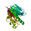
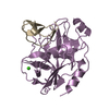
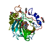
 Homo sapiens (human)
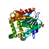
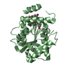
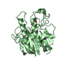
 X-RAY DIFFRACTION /
  Authors
 United States, 3items
  Citation
 Structure visualization
 Downloads & links
 Other downloads


 PDBj
PDBj


 Assembly


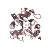
 Mass: 54.938 Da / Num. of mol.: 1 / Source method: obtained synthetically / Formula: Mn / Feature type: SUBJECT OF INVESTIGATION
 Mass: 24.305 Da / Num. of mol.: 2 / Source method: obtained synthetically / Formula: Mg / Feature type: SUBJECT OF INVESTIGATION
 Type: RNA linking / Mass: 404.161 Da / Num. of mol.: 1 / Source method: obtained synthetically / Formula: C9H14N2O12P2 / Feature type: SUBJECT OF INVESTIGATION / Comment: UDP*YM
 Mass: 122.143 Da / Num. of mol.: 1 / Source method: obtained synthetically / Formula: C4H12NO3 / Comment: pH buffer*YM
 Sample preparation
 Processing