 Movie
Movie Controller


+ Open data

- Basic information
| Entry | Database: PDB / ID: 6w6q | ||||||
|---|---|---|---|---|---|---|---|
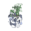
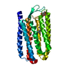
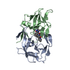
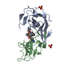
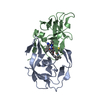
| Title | WT HTLV-1 Protease in Complex with Darunavir (DRV) | ||||||
 Components Components | HTLV-1 Protease | ||||||
 Keywords Keywords | HYDROLASE/HYDROLASE INHIBITOR / HTLV / PROTEASE / PROTEASE INHIBITOR / COMPLEX / HYDROLASE INHIBITOR COMPLEX / HYDROLASE / HYDROLASE-HYDROLASE INHIBITOR COMPLEX | ||||||
| Function / homology |  Function and homology information Function and homology information | ||||||
| Biological species |  Human T-cell leukemia virus type I Human T-cell leukemia virus type I | ||||||
| Method |  X-RAY DIFFRACTION / SYNCHROTRON / MOLECULAR REPLACEMENT / Resolution: 2.1 Å X-RAY DIFFRACTION / SYNCHROTRON / MOLECULAR REPLACEMENT / Resolution: 2.1 Å | ||||||
 Authors Authors | Lockbaum, G.J. / Henes, M. / Kosovrasti, K. / Nalivaika, E.A. / Ali, A. / KurtYilmaz, N. / Schiffer, C.A. | ||||||
| Funding support |  United States, 1items United States, 1items
| ||||||
 Citation Citation | Journal: To Be Published Title: To Be Determined Authors: Lockbaum, G.J. / Henes, M. / Kosovrasti, K. / Nalivaika, E.A. / Ali, A. / KurtYilmaz, N. / Schiffer, C.A. | ||||||
| History |
|
- Structure visualization
| Structure viewer | Molecule: MolmilJmol/JSmol |
|---|
- Downloads & links
-Download
| PDBx/mmCIF format | 6w6q.cif.gz | 62.6 KB | Display | PDBx/mmCIF format |
|---|---|---|---|---|
| PDB format | pdb6w6q.ent.gz | 44.8 KB | Display | PDB format |
| PDBx/mmJSON format | 6w6q.json.gz | Tree view | PDBx/mmJSON format | |
| Others |  Other downloads Other downloads |
-Validation report
| Arichive directory | https://data.pdbj.org/pub/pdb/validation_reports/w6/6w6qftp://data.pdbj.org/pub/pdb/validation_reports/w6/6w6q | HTTPS FTP |
|---|
-Related structure data
| Related structure data | 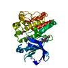 6auaC 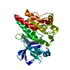 6aubC 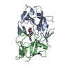 6w6rC 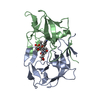 6w6sC 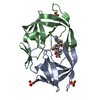 6w6tC  3wsjS S: Starting model for refinement C: citing same article ( |
|---|---|
| Similar structure data |


-Links
 PDBj
PDBj- Assembly
| Deposited unit | 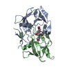
| ||||||||
|---|---|---|---|---|---|---|---|---|---|
| 1 |
| ||||||||
| Unit cell |
| ||||||||
| Components on special symmetry positions |
|
-Components
| #1: Protein | Mass: 12566.579 Da / Num. of mol.: 2 Source method: isolated from a genetically manipulated source Source: (gene. exp.) Human T-cell leukemia virus type I / Gene: pro / Details (production host): pXC35 / Production host:  #2: Chemical | ChemComp-017 / ( |   Mass: 547.664 Da / Num. of mol.: 1 / Source method: isolated from a natural source / Formula: C27H37N3O7S / Feature type: SUBJECT OF INVESTIGATION / Comment: medication, antiretroviral*YM Mass: 547.664 Da / Num. of mol.: 1 / Source method: isolated from a natural source / Formula: C27H37N3O7S / Feature type: SUBJECT OF INVESTIGATION / Comment: medication, antiretroviral*YM#3: Water | ChemComp-HOH / |  Mass: 18.015 Da / Num. of mol.: 52 / Source method: isolated from a natural source / Formula: H2O Mass: 18.015 Da / Num. of mol.: 52 / Source method: isolated from a natural source / Formula: H2OHas ligand of interest | Y | |
|---|
-Experimental details
-Experiment
| Experiment | Method: X-RAY DIFFRACTION / Number of used crystals: 1 |
|---|
- Sample preparation
| Crystal | Density Matthews: 2.84 Å3/Da / Density % sol: 56.71 % |
|---|---|
| Crystal grow | Temperature: 293 K / Method: vapor diffusion, hanging drop / pH: 4.2 Details: 39-41% (v/v) PEG 300, 0.1M Phosphate/Citrate Buffer pH 4.2 |
-Data collection
| Diffraction | Mean temperature: 100 K / Serial crystal experiment: N |
|---|---|
| Diffraction source | Source: SYNCHROTRON / Site: APS / Beamline: 23-ID-D / Wavelength: 1.07812 Å |
| Detector | Type: DECTRIS PILATUS3 6M / Detector: PIXEL / Date: Mar 20, 2019 |
| Radiation | Protocol: SINGLE WAVELENGTH / Monochromatic (M) / Laue (L): M / Scattering type: x-ray |
| Radiation wavelength | Wavelength: 1.07812 Å / Relative weight: 1 |
| Reflection | Resolution: 2.1→42.1 Å / Num. obs: 17823 / % possible obs: 99.84 % / Redundancy: 9.1 % / Biso Wilson estimate: 55.16 Å2 / CC1/2: 0.998 / CC star: 1 / Rmerge(I) obs: 0.09417 / Rpim(I) all: 0.03357 / Net I/σ(I): 12.61 |
| Reflection shell | Resolution: 2.1→2.175 Å / Rmerge(I) obs: 1.996 / Mean I/σ(I) obs: 1.16 / Num. unique obs: 16621 / CC1/2: 0.366 / Rpim(I) all: 0.6757 |
- Processing
| Software |
| |||||||||||||||||||||||||||||||||||
|---|---|---|---|---|---|---|---|---|---|---|---|---|---|---|---|---|---|---|---|---|---|---|---|---|---|---|---|---|---|---|---|---|---|---|---|---|
| Refinement | Method to determine structure: MOLECULAR REPLACEMENT Starting model: 3wsj Resolution: 2.1→42.052 Å / SU ML: 0.3 / Cross valid method: THROUGHOUT / σ(F): 1.34 / Phase error: 31.08 / Stereochemistry target values: ML
| |||||||||||||||||||||||||||||||||||
| Solvent computation | Shrinkage radii: 0.9 Å / VDW probe radii: 1.11 Å / Solvent model: FLAT BULK SOLVENT MODEL | |||||||||||||||||||||||||||||||||||
| Displacement parameters | Biso max: 167.65 Å2 / Biso mean: 70.5757 Å2 / Biso min: 33.64 Å2 | |||||||||||||||||||||||||||||||||||
| Refinement step | Cycle: final / Resolution: 2.1→42.052 Å
| |||||||||||||||||||||||||||||||||||
| Refine LS restraints |
| |||||||||||||||||||||||||||||||||||
| LS refinement shell | Refine-ID: X-RAY DIFFRACTION / Rfactor Rfree error: 0 / % reflection obs: 100 %
|
