 Movie
Movie Controller


+ Open data

- Basic information
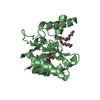
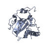
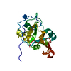
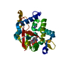
| Entry | Database: PDB / ID: 6vuv | ||||||
|---|---|---|---|---|---|---|---|
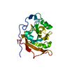
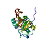
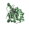
| Title | Scabin (S117A) toxin from Streptomyces scabies | ||||||
 Components Components | Scabin | ||||||
 Keywords Keywords | TOXIN / Transferase | ||||||
| Function / homology | : / : / Scabin-like / nucleotide binding / Putative secreted protein Function and homology information Function and homology information | ||||||
| Biological species |  Streptomyces scabiei (bacteria) Streptomyces scabiei (bacteria) | ||||||
| Method |  X-RAY DIFFRACTION / SYNCHROTRON / MOLECULAR REPLACEMENT / Resolution: 1.55 Å X-RAY DIFFRACTION / SYNCHROTRON / MOLECULAR REPLACEMENT / Resolution: 1.55 Å | ||||||
 Authors Authors | Lyons, B. / Lidster, T. / Merrill, A.R. | ||||||
| Funding support |  Canada, 1items Canada, 1items
| ||||||
 Citation Citation | Journal: Toxins / Year: 2021 Title: Mapping the DNA-Binding Motif of Scabin Toxin, a Guanine Modifying Enzyme from Streptomyces scabies . Authors: Vatta, M. / Lyons, B. / Heney, K.A. / Lidster, T. / Merrill, A.R. | ||||||
| History |
|
- Structure visualization
| Structure viewer | Molecule: MolmilJmol/JSmol |
|---|
- Downloads & links
-Download
| PDBx/mmCIF format | 6vuv.cif.gz | 76.2 KB | Display | PDBx/mmCIF format |
|---|---|---|---|---|
| PDB format | pdb6vuv.ent.gz | 56.3 KB | Display | PDB format |
| PDBx/mmJSON format | 6vuv.json.gz | Tree view | PDBx/mmJSON format | |
| Others |  Other downloads Other downloads |
-Validation report
| Arichive directory | https://data.pdbj.org/pub/pdb/validation_reports/vu/6vuvftp://data.pdbj.org/pub/pdb/validation_reports/vu/6vuv | HTTPS FTP |
|---|
-Related structure data
| Related structure data | 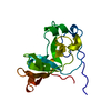 6vpaC  6vv4C  6vvfC  5dazS S: Starting model for refinement C: citing same article ( |
|---|---|
| Similar structure data |


-Links
 PDBj
PDBj- Assembly
| Deposited unit | 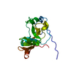
| ||||||||||||
|---|---|---|---|---|---|---|---|---|---|---|---|---|---|
| 1 |
| ||||||||||||
| Unit cell |
| ||||||||||||
| Components on special symmetry positions |
|
-Components
| #1: Protein | Mass: 21841.170 Da / Num. of mol.: 1 / Mutation: S117A Source method: isolated from a genetically manipulated source Source: (gene. exp.) Streptomyces scabiei (strain 87.22) (bacteria)Strain: 87.22 / Gene: SCAB_27771 / Production host: |
|---|---|
| #2: Water | ChemComp-HOH /  Mass: 18.015 Da / Num. of mol.: 151 / Source method: isolated from a natural source / Formula: H2O Mass: 18.015 Da / Num. of mol.: 151 / Source method: isolated from a natural source / Formula: H2O |
| Has protein modification | Y |
-Experimental details
-Experiment
| Experiment | Method: X-RAY DIFFRACTION / Number of used crystals: 1 |
|---|
- Sample preparation
| Crystal | Density Matthews: 2.31 Å3/Da / Density % sol: 46.64 % |
|---|---|
| Crystal grow | Temperature: 294 K / Method: vapor diffusion, hanging drop / pH: 6 Details: 100 mM potassium chloride, 50 mM sodium cacodylate trihydrate, pH 6.0, 16% PEG1000, 0.5 mM spermine |
-Data collection
| Diffraction | Mean temperature: 100 K / Serial crystal experiment: N |
|---|---|
| Diffraction source | Source: SYNCHROTRON / Site: CLSI / Beamline: 08ID-1 / Wavelength: 0.979 Å |
| Detector | Type: RAYONIX MX-300 / Detector: CCD / Date: Dec 13, 2016 |
| Radiation | Monochromator: double crystal Si(111) / Protocol: SINGLE WAVELENGTH / Monochromatic (M) / Laue (L): M / Scattering type: x-ray |
| Radiation wavelength | Wavelength: 0.979 Å / Relative weight: 1 |
| Reflection | Resolution: 1.55→43.61 Å / Num. obs: 28826 / % possible obs: 99.71 % / Redundancy: 4.2 % / CC1/2: 0.999 / Rmerge(I) obs: 0.04171 / Rpim(I) all: 0.02314 / Rrim(I) all: 0.04781 / Net I/σ(I): 17.72 |
| Reflection shell | Resolution: 1.55→1.605 Å / Redundancy: 4.1 % / Rmerge(I) obs: 0.9209 / Mean I/σ(I) obs: 1.64 / Num. unique obs: 2860 / CC1/2: 0.692 / Rpim(I) all: 0.5157 / % possible all: 99.62 |
- Processing
| Software |
| |||||||||||||||||||||||||||||||||||||||||||||||||||||||
|---|---|---|---|---|---|---|---|---|---|---|---|---|---|---|---|---|---|---|---|---|---|---|---|---|---|---|---|---|---|---|---|---|---|---|---|---|---|---|---|---|---|---|---|---|---|---|---|---|---|---|---|---|---|---|---|---|
| Refinement | Method to determine structure: MOLECULAR REPLACEMENT Starting model: PDB entry 5DAZ Resolution: 1.55→43.606 Å / SU ML: 0.18 / Cross valid method: THROUGHOUT / σ(F): 1.35 / Phase error: 23.7 / Stereochemistry target values: ML
| |||||||||||||||||||||||||||||||||||||||||||||||||||||||
| Solvent computation | Shrinkage radii: 0.9 Å / VDW probe radii: 1.11 Å / Solvent model: FLAT BULK SOLVENT MODEL | |||||||||||||||||||||||||||||||||||||||||||||||||||||||
| Displacement parameters | Biso max: 89.88 Å2 / Biso mean: 33.4009 Å2 / Biso min: 15.65 Å2 | |||||||||||||||||||||||||||||||||||||||||||||||||||||||
| Refinement step | Cycle: final / Resolution: 1.55→43.606 Å
| |||||||||||||||||||||||||||||||||||||||||||||||||||||||
| LS refinement shell | Refine-ID: X-RAY DIFFRACTION / Rfactor Rfree error: 0 / % reflection obs: 100 %
|
