 Movie
Movie Controller

[English] 日本語
 Yorodumi
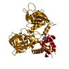
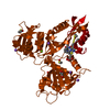
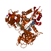
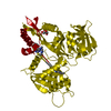
- PDB-6vur: Crystal structure of Eis from Mycobacterium tuberculosis in compl... -
+ Open data

- Basic information
| Entry | Database: PDB / ID: 6vur | ||||||
|---|---|---|---|---|---|---|---|
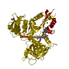
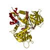
| Title | Crystal structure of Eis from Mycobacterium tuberculosis in complex with inhibitor SGT366 | ||||||
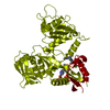
 Components Components | N-acetyltransferase Eis | ||||||
 Keywords Keywords | TRANSFERASE/TRANSFERASE INHIBITOR / Acetyltransferase / Resistance / Aminoglycoside / Competitive / TRANSFERASE / TRANSFERASE-TRANSFERASE INHIBITOR complex | ||||||
| Function / homology |  Function and homology information Function and homology informationeffector-mediated defense to host-produced reactive oxygen species / symbiont-mediated perturbation of host inflammatory response / symbiont-mediated perturbation of host innate immune response / Suppression of autophagy / symbiont-mediated suppression of host programmed cell death / aminoglycoside antibiotic catabolic process / aminoglycoside N-acetyltransferase activity / bacterial extracellular vesicle / symbiont-mediated perturbation of host programmed cell death / N-acetyltransferase activity ...effector-mediated defense to host-produced reactive oxygen species / symbiont-mediated perturbation of host inflammatory response / symbiont-mediated perturbation of host innate immune response / Suppression of autophagy / symbiont-mediated suppression of host programmed cell death / aminoglycoside antibiotic catabolic process / aminoglycoside N-acetyltransferase activity / bacterial extracellular vesicle / symbiont-mediated perturbation of host programmed cell death / N-acetyltransferase activity / biological process involved in interaction with host / host cell cytoplasmic vesicle / protein-lysine-acetyltransferase activity / Transferases; Acyltransferases; Transferring groups other than aminoacyl groups / host extracellular space / response to antibiotic / identical protein binding / cytosol Similarity search - Function | ||||||
| Biological species |   Mycobacterium tuberculosis (bacteria) Mycobacterium tuberculosis (bacteria) | ||||||
| Method |  X-RAY DIFFRACTION / SYNCHROTRON / MOLECULAR REPLACEMENT / Resolution: 2.2 Å X-RAY DIFFRACTION / SYNCHROTRON / MOLECULAR REPLACEMENT / Resolution: 2.2 Å | ||||||
 Authors Authors | Punetha, A. / Hou, C. / Ngo, H.X. / Garneau-Tsodikova, S. / Tsodikov, O.V. | ||||||
| Funding support |  United States, 1items United States, 1items
| ||||||
 Citation Citation | Journal: Acs Chem.Biol. / Year: 2020 Title: Structure-Guided Optimization of Inhibitors of Acetyltransferase Eis fromMycobacterium tuberculosis. Authors: Punetha, A. / Ngo, H.X. / Holbrook, S.Y.L. / Green, K.D. / Willby, M.J. / Bonnett, S.A. / Krieger, K. / Dennis, E.K. / Posey, J.E. / Parish, T. / Tsodikov, O.V. / Garneau-Tsodikova, S. | ||||||
| History |
|
- Structure visualization
| Structure viewer | Molecule: MolmilJmol/JSmol |
|---|
- Downloads & links
-Download
| PDBx/mmCIF format | 6vur.cif.gz | 101.1 KB | Display | PDBx/mmCIF format |
|---|---|---|---|---|
| PDB format | pdb6vur.ent.gz | 73.6 KB | Display | PDB format |
| PDBx/mmJSON format | 6vur.json.gz | Tree view | PDBx/mmJSON format | |
| Others |  Other downloads Other downloads |
-Validation report
| Summary document | 6vur_validation.pdf.gz | 363.1 KB | Display | wwPDB validaton report |
|---|---|---|---|---|
| Full document | 6vur_full_validation.pdf.gz | 363.4 KB | Display | |
| Data in XML | 6vur_validation.xml.gz | 2.1 KB | Display | |
| Data in CIF | 6vur_validation.cif.gz | 7.5 KB | Display | |
| Arichive directory | https://data.pdbj.org/pub/pdb/validation_reports/vu/6vurftp://data.pdbj.org/pub/pdb/validation_reports/vu/6vur | HTTPS FTP |
-Related structure data
| Related structure data |  6vusC  6vutC  6vuuC  6vuwC  6vuxC  6vuyC  6vuzC  6vv0C  6vv1C  6vv2C  6vv3C  3r1kS S: Starting model for refinement C: citing same article ( |
|---|---|
| Similar structure data |


-Links
 PDBj
PDBj


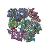
- Assembly
| Deposited unit | 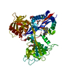
| ||||||||
|---|---|---|---|---|---|---|---|---|---|
| 1 | x 6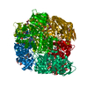
| ||||||||
| Unit cell |
|
-Components
-Protein , 1 types, 1 molecules A
| #1: Protein | Mass: 45999.113 Da / Num. of mol.: 1 / Mutation: C204A Source method: isolated from a genetically manipulated source Source: (gene. exp.) Mycobacterium tuberculosis (strain ATCC 25618 / H37Rv) (bacteria)Strain: ATCC 25618 / H37Rv / Gene: eis, Rv2416c, MTCY253.04 / Production host: References: UniProt: P9WFK7, Transferases; Acyltransferases; Transferring groups other than aminoacyl groups |
|---|
-Non-polymers , 7 types, 209 molecules 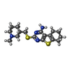


| #2: Chemical | ChemComp-ROG /  Mass: 348.529 Da / Num. of mol.: 1 / Source method: obtained synthetically / Formula: C17H24N4S2 / Feature type: SUBJECT OF INVESTIGATION Mass: 348.529 Da / Num. of mol.: 1 / Source method: obtained synthetically / Formula: C17H24N4S2 / Feature type: SUBJECT OF INVESTIGATION | ||||||||||
|---|---|---|---|---|---|---|---|---|---|---|---|
| #3: Chemical |  Mass: 96.063 Da / Num. of mol.: 3 / Source method: obtained synthetically / Formula: SO4 Mass: 96.063 Da / Num. of mol.: 3 / Source method: obtained synthetically / Formula: SO4#4: Chemical |  Mass: 92.094 Da / Num. of mol.: 3 / Source method: obtained synthetically / Formula: C3H8O3 Mass: 92.094 Da / Num. of mol.: 3 / Source method: obtained synthetically / Formula: C3H8O3#5: Chemical |  Mass: 106.120 Da / Num. of mol.: 2 / Source method: obtained synthetically / Formula: C4H10O3 Mass: 106.120 Da / Num. of mol.: 2 / Source method: obtained synthetically / Formula: C4H10O3#6: Chemical |  Mass: 22.990 Da / Num. of mol.: 2 / Source method: obtained synthetically / Formula: Na Mass: 22.990 Da / Num. of mol.: 2 / Source method: obtained synthetically / Formula: Na#7: Chemical |  Mass: 35.453 Da / Num. of mol.: 3 / Source method: obtained synthetically / Formula: Cl Mass: 35.453 Da / Num. of mol.: 3 / Source method: obtained synthetically / Formula: Cl#8: Water | ChemComp-HOH / | Mass: 18.015 Da / Num. of mol.: 195 / Source method: isolated from a natural source / Formula: H2O |
-Details
| Has ligand of interest | Y |
|---|
-Experimental details
-Experiment
| Experiment | Method: X-RAY DIFFRACTION / Number of used crystals: 1 |
|---|
- Sample preparation
| Crystal | Density Matthews: 3.94 Å3/Da / Density % sol: 68.77 % |
|---|---|
| Crystal grow | Temperature: 295 K / Method: vapor diffusion, hanging drop / pH: 8.5 Details: 100 mM Tris-HCl pH 8.5 adjusted at room temperature, 10% w/v PEG 8000, and 500 mM (NH4)2SO4 |
-Data collection
| Diffraction | Mean temperature: 100 K / Serial crystal experiment: N |
|---|---|
| Diffraction source | Source: SYNCHROTRON / Site: APS / Beamline: 22-ID / Wavelength: 1 Å |
| Detector | Type: DECTRIS EIGER X 16M / Detector: PIXEL / Date: Jul 11, 2018 |
| Radiation | Protocol: SINGLE WAVELENGTH / Monochromatic (M) / Laue (L): M / Scattering type: x-ray |
| Radiation wavelength | Wavelength: 1 Å / Relative weight: 1 |
| Reflection | Resolution: 2.2→50 Å / Num. obs: 36796 / % possible obs: 99.9 % / Redundancy: 7.4 % / CC1/2: 0.995 / Rmerge(I) obs: 0.164 / Net I/σ(I): 16.6 |
| Reflection shell | Resolution: 2.2→2.24 Å / Rmerge(I) obs: 1 / Num. unique obs: 1809 / CC1/2: 0.786 |
- Processing
| Software |
| ||||||||||||||||||||||||||||||||||||||||||||||||||||||||||||
|---|---|---|---|---|---|---|---|---|---|---|---|---|---|---|---|---|---|---|---|---|---|---|---|---|---|---|---|---|---|---|---|---|---|---|---|---|---|---|---|---|---|---|---|---|---|---|---|---|---|---|---|---|---|---|---|---|---|---|---|---|---|
| Refinement | Method to determine structure: MOLECULAR REPLACEMENT Starting model: 3R1K Resolution: 2.2→40 Å / Cor.coef. Fo:Fc: 0.958 / Cor.coef. Fo:Fc free: 0.943 / SU B: 5.129 / SU ML: 0.121 / Cross valid method: THROUGHOUT / σ(F): 0 / ESU R: 0.158 / ESU R Free: 0.145 / Stereochemistry target values: MAXIMUM LIKELIHOOD Details: HYDROGENS HAVE BEEN ADDED IN THE RIDING POSITIONS U VALUES : REFINED INDIVIDUALLY
| ||||||||||||||||||||||||||||||||||||||||||||||||||||||||||||
| Solvent computation | Ion probe radii: 0.8 Å / Shrinkage radii: 0.8 Å / VDW probe radii: 1.2 Å / Solvent model: MASK | ||||||||||||||||||||||||||||||||||||||||||||||||||||||||||||
| Displacement parameters | Biso max: 106.87 Å2 / Biso mean: 33.306 Å2 / Biso min: 18.6 Å2
| ||||||||||||||||||||||||||||||||||||||||||||||||||||||||||||
| Refinement step | Cycle: final / Resolution: 2.2→40 Å
| ||||||||||||||||||||||||||||||||||||||||||||||||||||||||||||
| Refine LS restraints |
| ||||||||||||||||||||||||||||||||||||||||||||||||||||||||||||
| LS refinement shell | Resolution: 2.2→2.257 Å / Rfactor Rfree error: 0 / Total num. of bins used: 20
|
