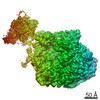
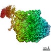
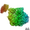
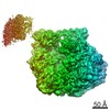
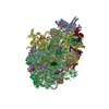
 Movie
Movie Controller

[English] 日本語
 Yorodumi
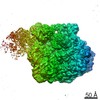
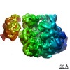
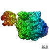
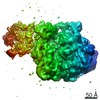
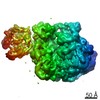
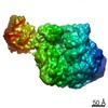
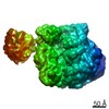
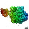
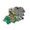
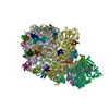
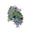
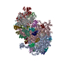
- PDB-6vu3: Cryo-EM structure of Escherichia coli transcription-translation c... -
+ Open data

- Basic information
| Entry | Database: PDB / ID: 6vu3 | |||||||||
|---|---|---|---|---|---|---|---|---|---|---|
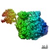
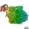
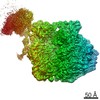
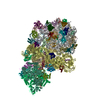
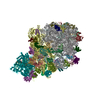
| Title | Cryo-EM structure of Escherichia coli transcription-translation complex A (TTC-A) containing mRNA with a 12 nt long spacer | |||||||||
 Components Components |
| |||||||||
 Keywords Keywords | RIBOSOME / bacterial coupled transcription-translation complex / TRANSCRIPTION-TRANSLATION complex | |||||||||
| Function / homology |  Function and homology information Function and homology informationtranscription elongation-coupled chromatin remodeling / DnaA-L2 complex / negative regulation of DNA-templated DNA replication initiation / regulation of DNA-templated transcription elongation / DNA-directed RNA polymerase complex / transcription antitermination / DNA-templated transcription termination / ribonucleoside binding / DNA-directed RNA polymerase / DNA-directed RNA polymerase activity ...transcription elongation-coupled chromatin remodeling / DnaA-L2 complex / negative regulation of DNA-templated DNA replication initiation / regulation of DNA-templated transcription elongation / DNA-directed RNA polymerase complex / transcription antitermination / DNA-templated transcription termination / ribonucleoside binding / DNA-directed RNA polymerase / DNA-directed RNA polymerase activity / large ribosomal subunit / transferase activity / ribosome biogenesis / 5S rRNA binding / ribosomal large subunit assembly / small ribosomal subunit / small ribosomal subunit rRNA binding / large ribosomal subunit rRNA binding / cytosolic small ribosomal subunit / cytosolic large ribosomal subunit / cytoplasmic translation / tRNA binding / protein dimerization activity / rRNA binding / structural constituent of ribosome / ribosome / translation / ribonucleoprotein complex / mRNA binding / DNA-templated transcription / magnesium ion binding / DNA binding / RNA binding / zinc ion binding / metal ion binding / cytoplasm / cytosol Similarity search - Function | |||||||||
| Biological species |  synthetic construct (others) | |||||||||
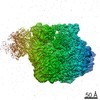
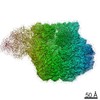
| Method | ELECTRON MICROSCOPY / single particle reconstruction / cryo EM / Resolution: 3.7 Å | |||||||||
 Authors Authors | Molodtsov, V. / Wang, C. / Su, M. / Ebright, R. | |||||||||
 Citation Citation | Journal: Science / Year: 2020 Title: Structural basis of transcription-translation coupling. Authors: Chengyuan Wang / Vadim Molodtsov / Emre Firlar / Jason T Kaelber / Gregor Blaha / Min Su / Richard H Ebright /  Abstract: In bacteria, transcription and translation are coupled processes in which the movement of RNA polymerase (RNAP)-synthesizing messenger RNA (mRNA) is coordinated with the movement of the first ...In bacteria, transcription and translation are coupled processes in which the movement of RNA polymerase (RNAP)-synthesizing messenger RNA (mRNA) is coordinated with the movement of the first ribosome-translating mRNA. Coupling is modulated by the transcription factors NusG (which is thought to bridge RNAP and the ribosome) and NusA. Here, we report cryo-electron microscopy structures of transcription-translation complexes (TTCs) containing different-length mRNA spacers between RNAP and the ribosome active-center P site. Structures of TTCs containing short spacers show a state incompatible with NusG bridging and NusA binding (TTC-A, previously termed "expressome"). Structures of TTCs containing longer spacers reveal a new state compatible with NusG bridging and NusA binding (TTC-B) and reveal how NusG bridges and NusA binds. We propose that TTC-B mediates NusG- and NusA-dependent transcription-translation coupling. | |||||||||
| History |
|
- Structure visualization
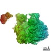
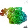
| Movie |
Movie viewer |
|---|---|
| Structure viewer | Molecule: MolmilJmol/JSmol |
- Downloads & links
-Download
| PDBx/mmCIF format | 6vu3.cif.gz | 6.7 MB | Display | PDBx/mmCIF format |
|---|---|---|---|---|
| PDB format | pdb6vu3.ent.gz | Display | PDB format | |
| PDBx/mmJSON format | 6vu3.json.gz | Tree view | PDBx/mmJSON format | |
| Others |  Other downloads Other downloads |
-Validation report
| Arichive directory | https://data.pdbj.org/pub/pdb/validation_reports/vu/6vu3ftp://data.pdbj.org/pub/pdb/validation_reports/vu/6vu3 | HTTPS FTP |
|---|
-Related structure data
| Related structure data | 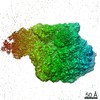 21386MC 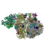 6vyqC 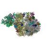 6vyrC 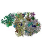 6vysC 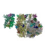 6vytC 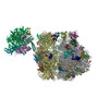 6vyuC  6vywC 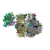 6vyxC 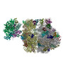 6vyyC 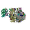 6vyzC 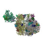 6vz2C 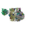 6vz3C 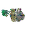 6vz5C 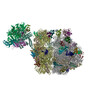 6vz7C 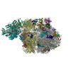 6vzjC 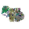 6x6tC 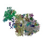 6x7fC 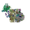 6x7kC 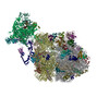 6x9qC 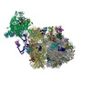 6xdqC 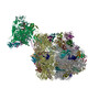 6xdrC  6xgfC 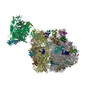 6xiiC 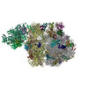 6xijC M: map data used to model this data C: citing same article ( |
|---|---|
| Similar structure data |


-Links
 PDBj
PDBj


- Assembly
| Deposited unit | 
|
|---|---|
| 1 |
|
-Components
+50S ribosomal protein ... , 32 types, 32 molecules 012349YZbcefghijklmnopqrstuvwxyz
-DNA chain , 2 types, 2 molecules 56
| #6: DNA chain | Mass: 11042.110 Da / Num. of mol.: 1 / Source method: obtained synthetically / Source: (synth.) synthetic construct (others) |
|---|---|
| #7: DNA chain | Mass: 8155.232 Da / Num. of mol.: 1 / Source method: obtained synthetically / Source: (synth.) synthetic construct (others) |
-RNA chain , 5 types, 6 molecules 7ABDad
| #8: RNA chain | Mass: 9238.331 Da / Num. of mol.: 1 / Source method: obtained synthetically / Source: (synth.) synthetic construct (others) | ||||||
|---|---|---|---|---|---|---|---|
| #10: RNA chain | Mass: 24496.617 Da / Num. of mol.: 2 / Source method: isolated from a natural source / Source: (natural) #16: RNA chain | | Mass: 499690.031 Da / Num. of mol.: 1 / Source method: isolated from a natural source / Source: (natural) #39: RNA chain | | Mass: 941635.438 Da / Num. of mol.: 1 / Source method: isolated from a natural source / Source: (natural) #42: RNA chain | | Mass: 38790.090 Da / Num. of mol.: 1 / Source method: isolated from a natural source / Source: (natural) |
-DNA-directed RNA polymerase subunit ... , 3 types, 4 molecules AAACADAE
| #11: Protein | Mass: 150820.875 Da / Num. of mol.: 1 / Source method: isolated from a natural source / Source: (natural) | ||
|---|---|---|---|
| #13: Protein | Mass: 25497.023 Da / Num. of mol.: 2 / Source method: isolated from a natural source / Source: (natural) References: UniProt: A0A5F0PYH4, DNA-directed RNA polymerase #14: Protein | | Mass: 155366.781 Da / Num. of mol.: 1 / Source method: isolated from a natural source / Source: (natural) References: UniProt: A0A4S1NBU2, DNA-directed RNA polymerase |
-Protein , 1 types, 1 molecules AB
| #12: Protein | Mass: 20560.523 Da / Num. of mol.: 1 / Source method: isolated from a natural source / Source: (natural) |
|---|
+30S ribosomal protein ... , 21 types, 21 molecules CEFGHIJKLMNOPQRSTUVWX
-Non-polymers , 2 types, 3 molecules 

| #65: Chemical | ChemComp-MG /  Mass: 24.305 Da / Num. of mol.: 1 / Source method: obtained synthetically / Formula: Mg Mass: 24.305 Da / Num. of mol.: 1 / Source method: obtained synthetically / Formula: Mg |
|---|---|
| #66: Chemical |  Mass: 65.409 Da / Num. of mol.: 2 / Source method: obtained synthetically / Formula: Zn Mass: 65.409 Da / Num. of mol.: 2 / Source method: obtained synthetically / Formula: Zn |
-Details
| Has ligand of interest | N |
|---|---|
| Has protein modification | Y |
-Experimental details
-Experiment
| Experiment | Method: ELECTRON MICROSCOPY |
|---|---|
| EM experiment | Aggregation state: PARTICLE / 3D reconstruction method: single particle reconstruction |
- Sample preparation
| Component | Name: Escherichia coli transcription-translation complex A (TTC-A) containing mRNA with a 12 nt long spacer Type: RIBOSOME / Entity ID: #1-#7, #9-#10, #12-#64 / Source: NATURAL |
|---|---|
| Molecular weight | Experimental value: NO |
| Source (natural) | Organism: |
| Buffer solution | pH: 7.5 |
| Specimen | Embedding applied: NO / Shadowing applied: NO / Staining applied: NO / Vitrification applied: YES |
| Vitrification | Cryogen name: ETHANE |
- Electron microscopy imaging
| Experimental equipment |  Model: Titan Krios / Image courtesy: FEI Company |
|---|---|
| Microscopy | Model: FEI TITAN KRIOS |
| Electron gun | Electron source:  FIELD EMISSION GUN / Accelerating voltage: 300 kV / Illumination mode: FLOOD BEAM FIELD EMISSION GUN / Accelerating voltage: 300 kV / Illumination mode: FLOOD BEAM |
| Electron lens | Mode: BRIGHT FIELD |
| Image recording | Electron dose: 45 e/Å2 / Film or detector model: GATAN K2 SUMMIT (4k x 4k) |
- Processing
| CTF correction | Type: PHASE FLIPPING AND AMPLITUDE CORRECTION |
|---|---|
| 3D reconstruction | Resolution: 3.7 Å / Resolution method: FSC 0.143 CUT-OFF / Num. of particles: 24959 / Symmetry type: POINT |
