 Movie
Movie Controller

[English] 日本語
 Yorodumi
- PDB-6vu0: CRYSTAL STRUCTURE OF THE C-TERMINAL DOMAIN OF ENZYME I OF THE BAC... -
+ Open data

- Basic information
| Entry | Database: PDB / ID: 6vu0 | ||||||
|---|---|---|---|---|---|---|---|
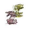
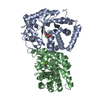
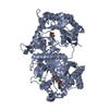
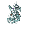
| Title | CRYSTAL STRUCTURE OF THE C-TERMINAL DOMAIN OF ENZYME I OF THE BACTERIAL PHOSPHOTRANSFERASE SYSTEM FROM THE ESCHERICHIA COLI ENZYME | ||||||
 Components Components | PEP-protein phosphotransferase system enzyme I | ||||||
 Keywords Keywords | TRANSFERASE / phosphoenolpyruvate-protein phosphotransferase PtsI / enzyme I | ||||||
| Function / homology |  Function and homology information Function and homology informationphosphoenolpyruvate-protein phosphotransferase / phosphoenolpyruvate-protein phosphotransferase activity / N-acetylglucosamine transport / phosphoenolpyruvate-dependent sugar phosphotransferase system / kinase activity / metal ion binding / identical protein binding / cytoplasm / cytosol Similarity search - Function | ||||||
| Biological species |  | ||||||
| Method |  X-RAY DIFFRACTION / SYNCHROTRON / MOLECULAR REPLACEMENT / Resolution: 3.5 Å X-RAY DIFFRACTION / SYNCHROTRON / MOLECULAR REPLACEMENT / Resolution: 3.5 Å | ||||||
 Authors Authors | Stewart Jr., C.E. | ||||||
| Funding support |  United States, 1items United States, 1items
| ||||||
 Citation Citation | Journal: J.Mol.Biol. / Year: 2020 Title: Hybrid Thermophilic/Mesophilic Enzymes Reveal a Role for Conformational Disorder in Regulation of Bacterial Enzyme I. Authors: Dotas, R.R. / Nguyen, T.T. / Stewart Jr., C.E. / Ghirlando, R. / Potoyan, D.A. / Venditti, V. | ||||||
| History |
|
- Structure visualization
| Structure viewer | Molecule: MolmilJmol/JSmol |
|---|
- Downloads & links
-Download
| PDBx/mmCIF format | 6vu0.cif.gz | 291.3 KB | Display | PDBx/mmCIF format |
|---|---|---|---|---|
| PDB format | pdb6vu0.ent.gz | 194.5 KB | Display | PDB format |
| PDBx/mmJSON format | 6vu0.json.gz | Tree view | PDBx/mmJSON format | |
| Others |  Other downloads Other downloads |
-Validation report
| Arichive directory | https://data.pdbj.org/pub/pdb/validation_reports/vu/6vu0ftp://data.pdbj.org/pub/pdb/validation_reports/vu/6vu0 | HTTPS FTP |
|---|
-Related structure data
| Related structure data |  6v9kSC  6vbjC S: Starting model for refinement C: citing same article ( |
|---|---|
| Similar structure data |

-Links
 PDBj
PDBj
- Assembly
| Deposited unit | 
| ||||||||||||||||||
|---|---|---|---|---|---|---|---|---|---|---|---|---|---|---|---|---|---|---|---|
| 1 |
| ||||||||||||||||||
| Unit cell |
| ||||||||||||||||||
| Noncrystallographic symmetry (NCS) | NCS domain:
NCS domain segments: Component-ID: 1 / Ens-ID: 1 / Beg auth comp-ID: ALA / Beg label comp-ID: ALA / End auth comp-ID: ILE / End label comp-ID: ILE / Auth seq-ID: 261 - 569 / Label seq-ID: 2 - 310
|
-Components
| #1: Protein | Mass: 35350.566 Da / Num. of mol.: 2 Source method: isolated from a genetically manipulated source Source: (gene. exp.) References: UniProt: A0A1V2SSS1, UniProt: P08839*PLUS, phosphoenolpyruvate-protein phosphotransferase #2: Chemical |   Mass: 96.063 Da / Num. of mol.: 2 / Source method: obtained synthetically / Formula: SO4 / Feature type: SUBJECT OF INVESTIGATION Mass: 96.063 Da / Num. of mol.: 2 / Source method: obtained synthetically / Formula: SO4 / Feature type: SUBJECT OF INVESTIGATIONHas ligand of interest | Y | |
|---|
-Experimental details
-Experiment
| Experiment | Method: X-RAY DIFFRACTION / Number of used crystals: 1 |
|---|
- Sample preparation
| Crystal | Density Matthews: 6.04 Å3/Da / Density % sol: 79.65 % |
|---|---|
| Crystal grow | Temperature: 292 K / Method: vapor diffusion, sitting drop / pH: 5.6 Details: 500 mM Ammonium sulfate, 1.00 M Lithium sulfate, 100 mM tri-Sodium citrate pH 5.6, 1.0 mM TEW |
-Data collection
| Diffraction | Mean temperature: 100 K / Serial crystal experiment: N | ||||||||||||||||||||||||||||||
|---|---|---|---|---|---|---|---|---|---|---|---|---|---|---|---|---|---|---|---|---|---|---|---|---|---|---|---|---|---|---|---|
| Diffraction source | Source: SYNCHROTRON / Site: APS / Beamline: 23-ID-D / Wavelength: 1.03322 Å | ||||||||||||||||||||||||||||||
| Detector | Type: DECTRIS PILATUS3 6M / Detector: PIXEL / Date: Oct 17, 2019 / Details: mirrors | ||||||||||||||||||||||||||||||
| Radiation | Monochromator: double crystal / Protocol: SINGLE WAVELENGTH / Monochromatic (M) / Laue (L): M / Scattering type: x-ray | ||||||||||||||||||||||||||||||
| Radiation wavelength | Wavelength: 1.03322 Å / Relative weight: 1 | ||||||||||||||||||||||||||||||
| Reflection | Resolution: 2.5→37.07 Å / Num. obs: 54988 / % possible obs: 91.1 % / Redundancy: 5.2 % / Biso Wilson estimate: 94.51 Å2 / CC1/2: 0.987 / Rmerge(I) obs: 0.45 / Rpim(I) all: 0.164 / Rrim(I) all: 0.483 / Net I/σ(I): 3 / Num. measured all: 285184 / Scaling rejects: 803 | ||||||||||||||||||||||||||||||
| Reflection shell | Diffraction-ID: 1
|
- Processing
| Software |
| ||||||||||||||||||||||||||||||||||||||||||||||||||||||||
|---|---|---|---|---|---|---|---|---|---|---|---|---|---|---|---|---|---|---|---|---|---|---|---|---|---|---|---|---|---|---|---|---|---|---|---|---|---|---|---|---|---|---|---|---|---|---|---|---|---|---|---|---|---|---|---|---|---|
| Refinement | Method to determine structure: MOLECULAR REPLACEMENT Starting model: 6V9K Resolution: 3.5→37.07 Å / SU ML: 0.5006 / Cross valid method: FREE R-VALUE / σ(F): 1.33 / Phase error: 30.5409 Stereochemistry target values: GeoStd + Monomer Library + CDL v1.2
| ||||||||||||||||||||||||||||||||||||||||||||||||||||||||
| Solvent computation | Shrinkage radii: 0.9 Å / VDW probe radii: 1.11 Å / Solvent model: FLAT BULK SOLVENT MODEL | ||||||||||||||||||||||||||||||||||||||||||||||||||||||||
| Displacement parameters | Biso mean: 109.7 Å2 | ||||||||||||||||||||||||||||||||||||||||||||||||||||||||
| Refinement step | Cycle: LAST / Resolution: 3.5→37.07 Å
| ||||||||||||||||||||||||||||||||||||||||||||||||||||||||
| Refine LS restraints |
| ||||||||||||||||||||||||||||||||||||||||||||||||||||||||
| LS refinement shell |
|
