 Movie
Movie Controller


+ Open data

- Basic information
| Entry | Database: PDB / ID: 6vsh | ||||||
|---|---|---|---|---|---|---|---|
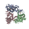
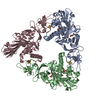
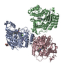
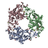
| Title | Crystal structure of apo Dicamba Monooxygenase | ||||||
 Components Components | Dicamba O-demethylase, oxygenase component | ||||||
 Keywords Keywords | OXIDOREDUCTASE / ELECTRON TRANSPORT | ||||||
| Function / homology |  Function and homology information Function and homology informationOxidoreductases; Acting on paired donors, with incorporation or reduction of molecular oxygen; With reduced iron-sulfur protein as one donor, and incorporation of one atom of oxygen into the other donor / monooxygenase activity / 2 iron, 2 sulfur cluster binding / metal ion binding Similarity search - Function | ||||||
| Biological species |  Stenotrophomonas maltophilia (bacteria) Stenotrophomonas maltophilia (bacteria) | ||||||
| Method |  X-RAY DIFFRACTION / MOLECULAR REPLACEMENT / Resolution: 3 Å X-RAY DIFFRACTION / MOLECULAR REPLACEMENT / Resolution: 3 Å | ||||||
 Authors Authors | Rydel, T.J. | ||||||
 Citation Citation | Journal: J. Mol. Biol. / Year: 2009 Title: Dicamba monooxygenase: structural insights into a dynamic Rieske oxygenase that catalyzes an exocyclic monooxygenation. Authors: D'Ordine, R.L. / Rydel, T.J. / Storek, M.J. / Sturman, E.J. / Moshiri, F. / Bartlett, R.K. / Brown, G.R. / Eilers, R.J. / Dart, C. / Qi, Y. / Flasinski, S. / Franklin, S. | ||||||
| History |
|
- Structure visualization
| Structure viewer | Molecule: MolmilJmol/JSmol |
|---|
- Downloads & links
-Download
| PDBx/mmCIF format | 6vsh.cif.gz | 243 KB | Display | PDBx/mmCIF format |
|---|---|---|---|---|
| PDB format | pdb6vsh.ent.gz | 157.9 KB | Display | PDB format |
| PDBx/mmJSON format | 6vsh.json.gz | Tree view | PDBx/mmJSON format | |
| Others |  Other downloads Other downloads |
-Validation report
| Arichive directory | https://data.pdbj.org/pub/pdb/validation_reports/vs/6vshftp://data.pdbj.org/pub/pdb/validation_reports/vs/6vsh | HTTPS FTP |
|---|
-Related structure data
| Related structure data |  3gb4C  3gobC  3gteSC  3gtsC S: Starting model for refinement C: citing same article ( |
|---|---|
| Similar structure data |


-Links
 PDBj
PDBj


- Assembly
| Deposited unit | 
| |||||||||||||||||||||||||||||||||||||||||||||||||||||||||||||||||||||||
|---|---|---|---|---|---|---|---|---|---|---|---|---|---|---|---|---|---|---|---|---|---|---|---|---|---|---|---|---|---|---|---|---|---|---|---|---|---|---|---|---|---|---|---|---|---|---|---|---|---|---|---|---|---|---|---|---|---|---|---|---|---|---|---|---|---|---|---|---|---|---|---|---|
| 1 |
| |||||||||||||||||||||||||||||||||||||||||||||||||||||||||||||||||||||||
| Unit cell |
| |||||||||||||||||||||||||||||||||||||||||||||||||||||||||||||||||||||||
| Noncrystallographic symmetry (NCS) | NCS domain:
NCS domain segments: Ens-ID: 1
|