 Movie
Movie Controller

[English] 日本語
 Yorodumi
- PDB-6vqp: Structure of CalU17 from the Calicheamicin Biosynthesis Pathway o... -
+ Open data

- Basic information
| Entry | Database: PDB / ID: 6vqp | ||||||||||||
|---|---|---|---|---|---|---|---|---|---|---|---|---|---|
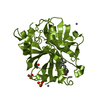
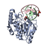
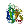
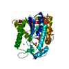
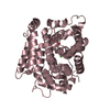
| Title | Structure of CalU17 from the Calicheamicin Biosynthesis Pathway of Micromonospora echinospora | ||||||||||||
 Components Components |
| ||||||||||||
 Keywords Keywords | UNKNOWN FUNCTION / Enediyne biosynthetic pathway / Calicheamicin Biosynthesis | ||||||||||||
| Function / homology | formylglycine-generating oxidase activity / : / Sulfatase-modifying factor enzyme / Sulfatase-modifying factor enzyme 1-like domain / Sulfatase-modifying factor enzyme superfamily / C-type lectin fold / metal ion binding / CalU17 Function and homology information Function and homology information | ||||||||||||
| Biological species |  Micromonospora echinospora (bacteria) Micromonospora echinospora (bacteria) | ||||||||||||
| Method |  X-RAY DIFFRACTION / SYNCHROTRON / MOLECULAR REPLACEMENT / molecular replacement / Resolution: 2 Å X-RAY DIFFRACTION / SYNCHROTRON / MOLECULAR REPLACEMENT / molecular replacement / Resolution: 2 Å | ||||||||||||
 Authors Authors | Kosgei, A.J. / Miller, M.D. / Xu, W. / Van Lanen, S.G. / Thorson, J.S. / Phillips Jr., G.N. | ||||||||||||
| Funding support |  United States, 3items United States, 3items
| ||||||||||||
 Citation Citation | Journal: To Be Published Title: Structure of DynF from the Dynemicin Biosynthesis Pathway of Micromonospora chersina Authors: Kosgei, A.J. / Miller, M.D. / Xu, W. / Van Lanen, S.G. / Thorson, J.S. / Phillips Jr., G.N. | ||||||||||||
| History |
|
- Structure visualization
| Structure viewer | Molecule: MolmilJmol/JSmol |
|---|
- Downloads & links
-Download
| PDBx/mmCIF format | 6vqp.cif.gz | 132.6 KB | Display | PDBx/mmCIF format |
|---|---|---|---|---|
| PDB format | pdb6vqp.ent.gz | 101.8 KB | Display | PDB format |
| PDBx/mmJSON format | 6vqp.json.gz | Tree view | PDBx/mmJSON format | |
| Others |  Other downloads Other downloads |
-Validation report
| Arichive directory | https://data.pdbj.org/pub/pdb/validation_reports/vq/6vqpftp://data.pdbj.org/pub/pdb/validation_reports/vq/6vqp | HTTPS FTP |
|---|
-Related structure data
| Related structure data | 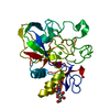 1y1eS S: Starting model for refinement |
|---|---|
| Similar structure data |


-Links
 PDBj
PDBj


- Assembly
| Deposited unit | 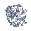
| |||||||||
|---|---|---|---|---|---|---|---|---|---|---|
| 1 |
| |||||||||
| Unit cell |
| |||||||||
| Components on special symmetry positions |
|
-Components
| #1: Protein | Mass: 37235.180 Da / Num. of mol.: 1 Source method: isolated from a genetically manipulated source Source: (gene. exp.) Micromonospora echinospora (bacteria) / Gene: calU17 / Production host: | ||||||
|---|---|---|---|---|---|---|---|
| #2: Protein/peptide | Mass: 2051.233 Da / Num. of mol.: 1 Source method: isolated from a genetically manipulated source Details: Chain Q, is the N-terminus tag from a neighboring chain. Due to the disorderd gap from residue -3 to 16, it is not clear which of two possible crystallographic symmetry transformations would ...Details: Chain Q, is the N-terminus tag from a neighboring chain. Due to the disorderd gap from residue -3 to 16, it is not clear which of two possible crystallographic symmetry transformations would transform chain Q to the position of the N-terminus of chain A. It is likely either Y+1/2, -X+1/2, Z+1/4 or -Y, -X+1, -Z+1/2 Source: (gene. exp.) Micromonospora echinospora (bacteria) / Gene: calU17 / Production host: | ||||||
| #3: Chemical |   Mass: 24.305 Da / Num. of mol.: 2 / Source method: obtained synthetically / Formula: Mg Mass: 24.305 Da / Num. of mol.: 2 / Source method: obtained synthetically / Formula: Mg#4: Chemical |   Mass: 92.094 Da / Num. of mol.: 2 / Source method: obtained synthetically / Formula: C3H8O3 Mass: 92.094 Da / Num. of mol.: 2 / Source method: obtained synthetically / Formula: C3H8O3#5: Water | ChemComp-HOH / |  Mass: 18.015 Da / Num. of mol.: 325 / Source method: isolated from a natural source / Formula: H2O Mass: 18.015 Da / Num. of mol.: 325 / Source method: isolated from a natural source / Formula: H2OHas ligand of interest | N | |
-Experimental details
-Experiment
| Experiment | Method: X-RAY DIFFRACTION / Number of used crystals: 1 |
|---|
- Sample preparation
| Crystal | Density Matthews: 2.03 Å3/Da / Density % sol: 39.33 % Description: It took a month fro the crystals to grow. The size of the crystals were about 70 microns length, width and height. |
|---|---|
| Crystal grow | Temperature: 277 K / Method: vapor diffusion, sitting drop / pH: 6.5 / Details: 0.1M Bis-Tris pH 6.5 20% w/v PEG 1500 Temp details: Set the crystallization trays at room temperature then incubated at 4 degrees |
-Data collection
| Diffraction | Mean temperature: 100 K / Serial crystal experiment: N | ||||||||||||||||||||||||||||||||||||||||||||||||||||||||||||||||||||||||||||||||||||||||||||||||||||
|---|---|---|---|---|---|---|---|---|---|---|---|---|---|---|---|---|---|---|---|---|---|---|---|---|---|---|---|---|---|---|---|---|---|---|---|---|---|---|---|---|---|---|---|---|---|---|---|---|---|---|---|---|---|---|---|---|---|---|---|---|---|---|---|---|---|---|---|---|---|---|---|---|---|---|---|---|---|---|---|---|---|---|---|---|---|---|---|---|---|---|---|---|---|---|---|---|---|---|---|---|---|
| Diffraction source | Source: SYNCHROTRON / Site: APS / Beamline: 23-ID-B / Wavelength: 1.0332 Å | ||||||||||||||||||||||||||||||||||||||||||||||||||||||||||||||||||||||||||||||||||||||||||||||||||||
| Detector | Type: DECTRIS EIGER X 16M / Detector: PIXEL / Date: Oct 16, 2018 | ||||||||||||||||||||||||||||||||||||||||||||||||||||||||||||||||||||||||||||||||||||||||||||||||||||
| Radiation | Monochromator: Double crystal cryo-cooled / Protocol: SINGLE WAVELENGTH / Monochromatic (M) / Laue (L): M / Scattering type: x-ray | ||||||||||||||||||||||||||||||||||||||||||||||||||||||||||||||||||||||||||||||||||||||||||||||||||||
| Radiation wavelength | Wavelength: 1.0332 Å / Relative weight: 1 | ||||||||||||||||||||||||||||||||||||||||||||||||||||||||||||||||||||||||||||||||||||||||||||||||||||
| Reflection | Resolution: 2→48.39 Å / Num. obs: 23026 / % possible obs: 99.8 % / Redundancy: 12.622 % / Biso Wilson estimate: 38.227 Å2 / CC1/2: 0.999 / Rmerge(I) obs: 0.106 / Rrim(I) all: 0.111 / Χ2: 1.06 / Net I/σ(I): 16.03 / Num. measured all: 290624 | ||||||||||||||||||||||||||||||||||||||||||||||||||||||||||||||||||||||||||||||||||||||||||||||||||||
| Reflection shell | Diffraction-ID: 1
|
-Phasing
| Phasing | Method: molecular replacement |
|---|
- Processing
| Software |
| |||||||||||||||||||||||||||||||||||||||||||||||||||||||||||||||
|---|---|---|---|---|---|---|---|---|---|---|---|---|---|---|---|---|---|---|---|---|---|---|---|---|---|---|---|---|---|---|---|---|---|---|---|---|---|---|---|---|---|---|---|---|---|---|---|---|---|---|---|---|---|---|---|---|---|---|---|---|---|---|---|---|
| Refinement | Method to determine structure: MOLECULAR REPLACEMENT Starting model: 1y1e Resolution: 2→48.39 Å / SU ML: 0.2 / Cross valid method: THROUGHOUT / σ(F): 1.35 / Phase error: 19.8 / Stereochemistry target values: ML
| |||||||||||||||||||||||||||||||||||||||||||||||||||||||||||||||
| Solvent computation | Shrinkage radii: 0.9 Å / VDW probe radii: 1.11 Å / Solvent model: FLAT BULK SOLVENT MODEL | |||||||||||||||||||||||||||||||||||||||||||||||||||||||||||||||
| Displacement parameters | Biso max: 151.11 Å2 / Biso mean: 42.2929 Å2 / Biso min: 22.66 Å2 | |||||||||||||||||||||||||||||||||||||||||||||||||||||||||||||||
| Refinement step | Cycle: final / Resolution: 2→48.39 Å
| |||||||||||||||||||||||||||||||||||||||||||||||||||||||||||||||
| LS refinement shell | Refine-ID: X-RAY DIFFRACTION / Rfactor Rfree error: 0 / Total num. of bins used: 8
|
