 Movie
Movie Controller

[English] 日本語
 Yorodumi
- PDB-6val: Cryo-EM structure of an undecameric chicken CALHM1 and human CALH... -
+ Open data

- Basic information
| Entry | Database: PDB / ID: 6val | |||||||||
|---|---|---|---|---|---|---|---|---|---|---|
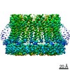
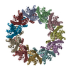
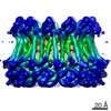
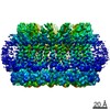
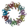
| Title | Cryo-EM structure of an undecameric chicken CALHM1 and human CALHM2 chimera | |||||||||
 Components Components | Green fluorescent protein, CALHM1,CALMH2 chimera | |||||||||
 Keywords Keywords | MEMBRANE PROTEIN / taste / assembly / calcium / chimera | |||||||||
| Function / homology |  Function and homology information Function and homology informationregulation of microglial cell activation / ATP export / voltage-gated monoatomic ion channel activity / calcium ion import / calcium-activated cation channel activity / monoatomic cation channel activity / bioluminescence / generation of precursor metabolites and energy / regulation of synaptic plasticity / positive regulation of apoptotic process / plasma membrane Similarity search - Function | |||||||||
| Biological species |   Aequorea victoria (jellyfish) Aequorea victoria (jellyfish)  Homo sapiens (human) Homo sapiens (human) | |||||||||
| Method | ELECTRON MICROSCOPY / single particle reconstruction / cryo EM / Resolution: 3.87 Å | |||||||||
 Authors Authors | Syrjanen, J.L. / Chou, T.H. / Furukawa, H. | |||||||||
| Funding support |  United States, 2items United States, 2items
| |||||||||
 Citation Citation | Journal: Nat Struct Mol Biol / Year: 2020 Title: Structure and assembly of calcium homeostasis modulator proteins. Authors: Johanna L Syrjanen / Kevin Michalski / Tsung-Han Chou / Timothy Grant / Shanlin Rao / Noriko Simorowski / Stephen J Tucker / Nikolaus Grigorieff / Hiro Furukawa /  Abstract: The biological membranes of many cell types contain large-pore channels through which a wide variety of ions and metabolites permeate. Examples include connexin, innexin and pannexin, which form gap ...The biological membranes of many cell types contain large-pore channels through which a wide variety of ions and metabolites permeate. Examples include connexin, innexin and pannexin, which form gap junctions and/or bona fide cell surface channels. The most recently identified large-pore channels are the calcium homeostasis modulators (CALHMs), through which ions and ATP permeate in a voltage-dependent manner to control neuronal excitability, taste signaling and pathologies of depression and Alzheimer's disease. Despite such critical biological roles, the structures and patterns of their oligomeric assembly remain unclear. Here, we reveal the structures of two CALHMs, chicken CALHM1 and human CALHM2, by single-particle cryo-electron microscopy (cryo-EM), which show novel assembly of the four transmembrane helices into channels of octamers and undecamers, respectively. Furthermore, molecular dynamics simulations suggest that lipids can favorably assemble into a bilayer within the larger CALHM2 pore, but not within CALHM1, demonstrating the potential correlation between pore size, lipid accommodation and channel activity. | |||||||||
| History |
|
- Structure visualization
| Movie |
Movie viewer |
|---|---|
| Structure viewer | Molecule: MolmilJmol/JSmol |
- Downloads & links
-Download
| PDBx/mmCIF format | 6val.cif.gz | 526.6 KB | Display | PDBx/mmCIF format |
|---|---|---|---|---|
| PDB format | pdb6val.ent.gz | 404.1 KB | Display | PDB format |
| PDBx/mmJSON format | 6val.json.gz | Tree view | PDBx/mmJSON format | |
| Others |  Other downloads Other downloads |
-Validation report
| Arichive directory | https://data.pdbj.org/pub/pdb/validation_reports/va/6valftp://data.pdbj.org/pub/pdb/validation_reports/va/6val | HTTPS FTP |
|---|
-Related structure data
| Related structure data | 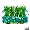 21142MC  6vaiC  6vakC  6vamC M: map data used to model this data C: citing same article ( |
|---|---|
| Similar structure data | |
| EM raw data | EMPIAR-10486 (Title: Chicken CALHM1 - Human CALHM2 chimera / Data size: 1.1 TB Data #1: Unaligned movies (.tif) for chicken CALHM1 - human CALHM2 chimera in nanodisc [micrographs - multiframe]) |


-Links
 PDBj
PDBj

- Assembly
| Deposited unit | 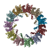
|
|---|---|
| 1 |
|
-Components
| #1: Protein | Mass: 69023.617 Da / Num. of mol.: 11 Fragment: GFP + CALHM1 (UNP residues 2-204) + CALMH2 (UNP residues 207-323) Source method: isolated from a genetically manipulated source Source: (gene. exp.) Aequorea victoria (jellyfish), (gene. exp.) Homo sapiens (human)Gene: GFP, CALHM1, CALHM2, FAM26B / Production host:   Spodoptera frugiperda (fall armyworm) Spodoptera frugiperda (fall armyworm)References: UniProt: P42212, UniProt: A0A1D5NWS1, UniProt: Q9HA72 Has protein modification | Y | |
|---|
-Experimental details
-Experiment
| Experiment | Method: ELECTRON MICROSCOPY |
|---|---|
| EM experiment | Aggregation state: PARTICLE / 3D reconstruction method: single particle reconstruction |
- Sample preparation
| Component | Name: chimera of chicken CALHM1 and human CALHM2 / Type: COMPLEX / Entity ID: all / Source: RECOMBINANT |
|---|---|
| Molecular weight | Experimental value: NO |
| Source (natural) | Organism: Homo sapiens (human) |
| Source (recombinant) | Organism: Spodoptera frugiperda (fall armyworm) |
| Buffer solution | pH: 7.5 |
| Specimen | Embedding applied: NO / Shadowing applied: NO / Staining applied: NO / Vitrification applied: YES |
| Specimen support | Details: unspecified |
| Vitrification | Instrument: FEI VITROBOT MARK IV / Cryogen name: ETHANE / Humidity: 85 % / Chamber temperature: 288.15 K / Details: Blot for 4 sec before plunging |
- Electron microscopy imaging
| Experimental equipment |  Model: Titan Krios / Image courtesy: FEI Company |
|---|---|
| Microscopy | Model: FEI TITAN KRIOS |
| Electron gun | Electron source:  FIELD EMISSION GUN / Accelerating voltage: 300 kV / Illumination mode: FLOOD BEAM FIELD EMISSION GUN / Accelerating voltage: 300 kV / Illumination mode: FLOOD BEAM |
| Electron lens | Mode: BRIGHT FIELD |
| Image recording | Electron dose: 57.2 e/Å2 / Film or detector model: GATAN K2 SUMMIT (4k x 4k) |
- Processing
| EM software |
| ||||||||||||
|---|---|---|---|---|---|---|---|---|---|---|---|---|---|
| CTF correction | Type: PHASE FLIPPING AND AMPLITUDE CORRECTION | ||||||||||||
| 3D reconstruction | Resolution: 3.87 Å / Resolution method: FSC 0.143 CUT-OFF / Num. of particles: 123664 / Symmetry type: POINT |
