| Entry | Database: PDB / ID: 6v6h
|
|---|
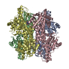
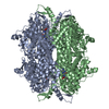
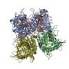
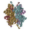
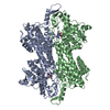
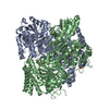
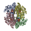
| Title | Crystal structure of histidine ammonia-lyase from Trypanosoma cruzi |
|---|
 Components Components | Histidine ammonia-lyase |
|---|
 Keywords Keywords | LYASE / histidine ammonia-lyase / protection domain absence |
|---|
| Function / homology |  Function and homology information Function and homology information
Histidine ammonia-lyase / Phenylalanine/histidine ammonia-lyases, active site / Phenylalanine and histidine ammonia-lyases signature. / Aromatic amino acid lyase / Aromatic amino acid lyase / Fumarase/histidase, N-terminal / L-Aspartase-likeSimilarity search - Domain/homology |
|---|
| Biological species |   Trypanosoma cruzi (eukaryote) Trypanosoma cruzi (eukaryote) |
|---|
| Method |  X-RAY DIFFRACTION / SYNCHROTRON / MOLECULAR REPLACEMENT / Resolution: 2.55 Å X-RAY DIFFRACTION / SYNCHROTRON / MOLECULAR REPLACEMENT / Resolution: 2.55 Å |
|---|
 Authors Authors | Miranda, R.R. / Silva, M. / Barison, M.J. / Silber, A.M. / Iulek, J. |
|---|
| Funding support | Belize, 1items | Organization | Grant number | Country |
|---|
| Brazilian National Council for Scientific and Technological Development | 573607/2008-7 and 08/57910-0 | Belize |
|
|---|
 Citation Citation | Journal: Biochimie / Year: 2020
Title: Crystal structure of histidine ammonia-lyase from Trypanosoma cruzi.
Authors: Miranda, R.R. / Silva, M. / Barison, M.J. / Silber, A.M. / Iulek, J. |
|---|
| History | | Deposition | Dec 5, 2019 | Deposition site: RCSB / Processing site: RCSB |
|---|
| Revision 1.0 | Jun 10, 2020 | Provider: repository / Type: Initial release |
|---|
| Revision 1.1 | Jul 15, 2020 | Group: Database references / Category: citation
Item: _citation.journal_volume / _citation.page_first / _citation.page_last |
|---|
| Revision 1.2 | Oct 11, 2023 | Group: Advisory / Data collection ...Advisory / Data collection / Database references / Derived calculations / Refinement description
Category: chem_comp_atom / chem_comp_bond ...chem_comp_atom / chem_comp_bond / database_2 / pdbx_initial_refinement_model / pdbx_unobs_or_zero_occ_atoms / struct_conn
Item: _database_2.pdbx_DOI / _database_2.pdbx_database_accession / _struct_conn.pdbx_leaving_atom_flag |
|---|
| Revision 2.0 | Nov 15, 2023 | Group: Atomic model / Data collection / Derived calculations
Category: atom_site / atom_site_anisotrop ...atom_site / atom_site_anisotrop / chem_comp_atom / chem_comp_bond / pdbx_validate_main_chain_plane / pdbx_validate_peptide_omega / pdbx_validate_rmsd_angle / pdbx_validate_torsion / struct_conn
Item: _atom_site.auth_atom_id / _atom_site.label_atom_id ..._atom_site.auth_atom_id / _atom_site.label_atom_id / _atom_site_anisotrop.pdbx_auth_atom_id / _atom_site_anisotrop.pdbx_label_atom_id / _chem_comp_atom.atom_id / _chem_comp_bond.atom_id_1 / _chem_comp_bond.atom_id_2 / _struct_conn.ptnr1_label_atom_id / _struct_conn.ptnr2_label_atom_id |
|---|
| Revision 2.1 | Nov 13, 2024 | Group: Structure summary / Category: pdbx_entry_details / pdbx_modification_feature / Item: _pdbx_entry_details.has_protein_modification |
|---|
| Revision 3.0 | Mar 18, 2026 | Group: Polymer sequence / Category: entity_poly / Item: _entity_poly.pdbx_seq_one_letter_code_can |
|---|
|
|---|
 Movie
Movie Controller

 Yorodumi
 Open data
 Basic information
 Structure visualization
 Downloads & links
 Other downloads


 PDBj
PDBj

 Assembly