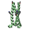
 Movie
Movie Controller


+ Open data

- Basic information
| Entry | Database: PDB / ID: 6v50 | ||||||
|---|---|---|---|---|---|---|---|
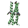
| Title | Coiled-coil Trimer with Glu:Ser:Lys Triad with K7A mutation | ||||||
 Components Components | Coiled-coil Trimer with Glu:Ser:Lys Triad with K7A mutation | ||||||
 Keywords Keywords | DE NOVO PROTEIN / Trimer / Helix | ||||||
| Biological species | synthetic construct (others) | ||||||
| Method |  X-RAY DIFFRACTION / MOLECULAR REPLACEMENT / Resolution: 2.285 Å X-RAY DIFFRACTION / MOLECULAR REPLACEMENT / Resolution: 2.285 Å | ||||||
 Authors Authors | Smith, M.S. / Stern, K.L. / Billings, W.M. / Price, J.L. | ||||||
| Funding support |  United States, 1items United States, 1items
| ||||||
 Citation Citation | Journal: Biochemistry / Year: 2020 Title: Context-Dependent Stabilizing Interactions among Solvent-Exposed Residues along the Surface of a Trimeric Helix Bundle. Authors: Stern, K.L. / Smith, M.S. / Billings, W.M. / Loftus, T.J. / Conover, B.M. / Della Corte, D. / Price, J.L. | ||||||
| History |
|
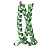
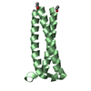
- Structure visualization
| Structure viewer | Molecule:  MolmilJmol/JSmol MolmilJmol/JSmol |
|---|
- Downloads & links
-Download
| PDBx/mmCIF format | 6v50.cif.gz | 23.4 KB | Display | PDBx/mmCIF format |
|---|---|---|---|---|
| PDB format | pdb6v50.ent.gz | 14.5 KB | Display | PDB format |
| PDBx/mmJSON format | 6v50.json.gz | Tree view | PDBx/mmJSON format | |
| Others |  Other downloads Other downloads |
-Validation report
| Summary document | 6v50_validation.pdf.gz | 248.7 KB | Display | wwPDB validaton report |
|---|---|---|---|---|
| Full document | 6v50_full_validation.pdf.gz | 248.7 KB | Display | |
| Data in XML | 6v50_validation.xml.gz | 1 KB | Display | |
| Data in CIF | 6v50_validation.cif.gz | 1.8 KB | Display | |
| Arichive directory | https://data.pdbj.org/pub/pdb/validation_reports/v5/6v50ftp://data.pdbj.org/pub/pdb/validation_reports/v5/6v50 | HTTPS FTP |
-Related structure data
| Related structure data | 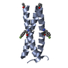 6os8C 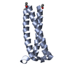 6osdC 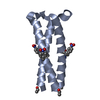 6ov9C 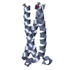 6ovsSC 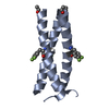 6ovuC 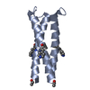 6ovvC  6q1wC 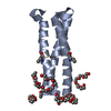 6q22C 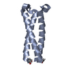 6q25C 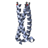 6u47C 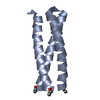 6v4yC 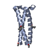 6v57C 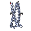 6v58C 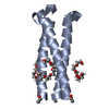 6v5gC 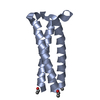 6v5iC 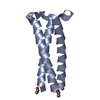 6v5jC S: Starting model for refinement C: citing same article ( |
|---|---|
| Similar structure data |


-Links
 PDBj
PDBj
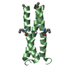
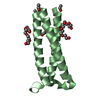
- Assembly
| Deposited unit | 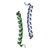
| ||||||||
|---|---|---|---|---|---|---|---|---|---|
| 1 | 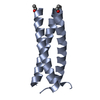
| ||||||||
| 2 | 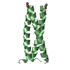
| ||||||||
| Unit cell |
|
-Components
| #1: Protein/peptide | Mass: 3568.031 Da / Num. of mol.: 2 / Source method: obtained synthetically / Source: (synth.) synthetic construct (others) #2: Water | ChemComp-HOH / |  Mass: 18.015 Da / Num. of mol.: 16 / Source method: isolated from a natural source / Formula: H2O Mass: 18.015 Da / Num. of mol.: 16 / Source method: isolated from a natural source / Formula: H2OHas ligand of interest | N | Has protein modification | Y | |
|---|
-Experimental details
-Experiment
| Experiment | Method: X-RAY DIFFRACTION / Number of used crystals: 1 |
|---|
- Sample preparation
| Crystal | Density Matthews: 2.04 Å3/Da / Density % sol: 39.77 % |
|---|---|
| Crystal grow | Temperature: 298 K / Method: vapor diffusion, sitting drop Details: 40% PEG300, 100 mM HEPES/NaOH, pH 7.5, 200 mM sodium chloride |
-Data collection
| Diffraction | Mean temperature: 100 K / Serial crystal experiment: N |
|---|---|
| Diffraction source | Source: ROTATING ANODE / Type: ENRAF-NONIUS FR591 / Wavelength: 1.5406 Å |
| Detector | Type: APEX II CCD / Detector: CCD / Date: Apr 2, 2019 |
| Radiation | Protocol: SINGLE WAVELENGTH / Monochromatic (M) / Laue (L): M / Scattering type: x-ray |
| Radiation wavelength | Wavelength: 1.5406 Å / Relative weight: 1 |
| Reflection | Resolution: 2.285→19.946 Å / Num. obs: 5113 / % possible obs: 99.9 % / Redundancy: 12.6 % / CC1/2: 0.999 / Rmerge(I) obs: 0.1 / Rpim(I) all: 0.029 / Rrim(I) all: 0.104 / Net I/σ(I): 12 |
| Reflection shell | Resolution: 2.285→2.615 Å / Rmerge(I) obs: 0.354 / Mean I/σ(I) obs: 2.3 / Num. unique obs: 258 / CC1/2: 0.946 / Rpim(I) all: 0.135 / Rrim(I) all: 0.104 |
- Processing
| Software |
| ||||||||||||||||||||||||
|---|---|---|---|---|---|---|---|---|---|---|---|---|---|---|---|---|---|---|---|---|---|---|---|---|---|
| Refinement | Method to determine structure: MOLECULAR REPLACEMENT Starting model: PDB entry 6OVS Resolution: 2.285→19.946 Å / SU ML: 0.17 / Cross valid method: THROUGHOUT / σ(F): 1.96 / Phase error: 28.79
| ||||||||||||||||||||||||
| Solvent computation | Shrinkage radii: 0.9 Å / VDW probe radii: 1.11 Å | ||||||||||||||||||||||||
| Displacement parameters | Biso max: 61.48 Å2 / Biso mean: 27.252 Å2 / Biso min: 11.71 Å2 | ||||||||||||||||||||||||
| Refinement step | Cycle: final / Resolution: 2.285→19.946 Å
| ||||||||||||||||||||||||
| Refine LS restraints |
| ||||||||||||||||||||||||
| LS refinement shell | Refine-ID: X-RAY DIFFRACTION / Rfactor Rfree error: 0
|
