 Movie
Movie Controller

[English] 日本語
 Yorodumi
- PDB-6v2n: Crystal structure of E. coli phosphoenolpyruvate carboxykinase mu... -
+ Open data

- Basic information
| Entry | Database: PDB / ID: 6v2n | |||||||||
|---|---|---|---|---|---|---|---|---|---|---|
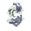
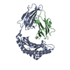
| Title | Crystal structure of E. coli phosphoenolpyruvate carboxykinase mutant Lys254Ser | |||||||||
 Components Components | Phosphoenolpyruvate carboxykinase (ATP) | |||||||||
 Keywords Keywords | LYASE / enzyme / Pepcarboxykinase | |||||||||
| Function / homology |  Function and homology information Function and homology informationphosphoenolpyruvate carboxykinase (ATP) / phosphoenolpyruvate carboxykinase (ATP) activity / gluconeogenesis / kinase activity / calcium ion binding / magnesium ion binding / ATP binding / metal ion binding / cytosol / cytoplasm Similarity search - Function | |||||||||
| Biological species |  | |||||||||
| Method |  X-RAY DIFFRACTION / SYNCHROTRON / MOLECULAR REPLACEMENT / Resolution: 1.65 Å X-RAY DIFFRACTION / SYNCHROTRON / MOLECULAR REPLACEMENT / Resolution: 1.65 Å | |||||||||
 Authors Authors | Sokaribo, A.S. / Cotelesage, J.H. / Novakovski, B. / Goldie, H. / Sanders, D. | |||||||||
 Citation Citation | Journal: Biochim Biophys Acta Gen Subj / Year: 2020 Title: Kinetic and structural analysis of Escherichia coli phosphoenolpyruvate carboxykinase mutants. Authors: Sokaribo, A. / Novakovski, B.A.A. / Cotelesage, J. / White, A.P. / Sanders, D. / Goldie, H. | |||||||||
| History |
|
- Structure visualization
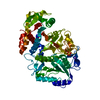
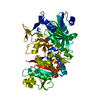
| Structure viewer | Molecule: MolmilJmol/JSmol |
|---|
- Downloads & links
-Download
| PDBx/mmCIF format | 6v2n.cif.gz | 233.9 KB | Display | PDBx/mmCIF format |
|---|---|---|---|---|
| PDB format | pdb6v2n.ent.gz | 183.2 KB | Display | PDB format |
| PDBx/mmJSON format | 6v2n.json.gz | Tree view | PDBx/mmJSON format | |
| Others |  Other downloads Other downloads |
-Validation report
| Arichive directory | https://data.pdbj.org/pub/pdb/validation_reports/v2/6v2nftp://data.pdbj.org/pub/pdb/validation_reports/v2/6v2n | HTTPS FTP |
|---|
-Related structure data
| Related structure data | 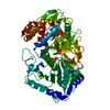 6comC 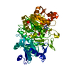 6v2lC 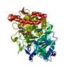 6v2mC 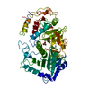 1oenS S: Starting model for refinement C: citing same article ( |
|---|---|
| Similar structure data |


-Links
 PDBj
PDBj- Assembly
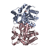
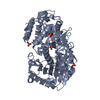
| Deposited unit | 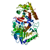
| ||||||||
|---|---|---|---|---|---|---|---|---|---|
| 1 |
| ||||||||
| Unit cell |
|
-Components
| #1: Protein | Mass: 59432.723 Da / Num. of mol.: 1 Source method: isolated from a genetically manipulated source Source: (gene. exp.) References: UniProt: A0A400L9R1, UniProt: P22259*PLUS, phosphoenolpyruvate carboxykinase (ATP) |
|---|---|
| #2: Chemical | ChemComp-CA /   Mass: 40.078 Da / Num. of mol.: 1 / Source method: obtained synthetically / Formula: Ca Mass: 40.078 Da / Num. of mol.: 1 / Source method: obtained synthetically / Formula: Ca |
| #3: Chemical | ChemComp-ACT /   Mass: 59.044 Da / Num. of mol.: 1 / Source method: obtained synthetically / Formula: C2H3O2 Mass: 59.044 Da / Num. of mol.: 1 / Source method: obtained synthetically / Formula: C2H3O2 |
| #4: Water | ChemComp-HOH /  Mass: 18.015 Da / Num. of mol.: 523 / Source method: isolated from a natural source / Formula: H2O Mass: 18.015 Da / Num. of mol.: 523 / Source method: isolated from a natural source / Formula: H2O |
| Has ligand of interest | N |
-Experimental details
-Experiment
| Experiment | Method: X-RAY DIFFRACTION / Number of used crystals: 1 |
|---|
- Sample preparation
| Crystal | Density Matthews: 2.39 Å3/Da / Density % sol: 48.64 % |
|---|---|
| Crystal grow | Temperature: 293 K / Method: microbatch Details: 2 ul drop containing 2 mg/ml Lys254Ser E. coli PCK, 5 mM MnCl2, 5mM MgCl2, 2mM ATP, 2mM pyruvate, 1 mM EDTA, 200 mM ammonium acetate, 100 mM sodium acetate pH 4.8, 0.01 mM DTT and 10% PEG ...Details: 2 ul drop containing 2 mg/ml Lys254Ser E. coli PCK, 5 mM MnCl2, 5mM MgCl2, 2mM ATP, 2mM pyruvate, 1 mM EDTA, 200 mM ammonium acetate, 100 mM sodium acetate pH 4.8, 0.01 mM DTT and 10% PEG 4000, was added to 2 ul drop containing 0.2 M calcium chloride and 20% PEG. Rod like crystals formed after 7 days, were harvested, and soaked in cryoprotectant solution (30% glycerol, 1mM EDTA, 100 mM sodium acetate, 200 mM ammonium acetate and 12% PEG 4000) for 10 seconds and flash cooled in liquid nitrogen |
-Data collection
| Diffraction | Mean temperature: 105 K / Serial crystal experiment: N |
|---|---|
| Diffraction source | Source: SYNCHROTRON / Site: CLSI  / Beamline: 08ID-1 / Wavelength: 0.9793 Å / Beamline: 08ID-1 / Wavelength: 0.9793 Å |
| Detector | Type: RAYONIX MX300HE / Detector: CCD / Date: Jul 10, 2014 |
| Radiation | Protocol: SINGLE WAVELENGTH / Monochromatic (M) / Laue (L): M / Scattering type: x-ray |
| Radiation wavelength | Wavelength: 0.9793 Å / Relative weight: 1 |
| Reflection | Resolution: 1.65→44.45 Å / Num. obs: 64733 / % possible obs: 99.83 % / Redundancy: 5.1 % / Biso Wilson estimate: 13.82 Å2 / CC1/2: 0.997 / Rmerge(I) obs: 0.07574 / Rpim(I) all: 0.03608 / Rrim(I) all: 0.08409 / Net I/σ(I): 14.67 |
| Reflection shell | Resolution: 1.65→1.709 Å / Redundancy: 4.9 % / Rmerge(I) obs: 0.3185 / Mean I/σ(I) obs: 4.56 / Num. unique obs: 6479 / CC1/2: 0.953 / Rpim(I) all: 0.1576 / Rrim(I) all: 0.3562 / % possible all: 99.92 |
- Processing
| Software |
| |||||||||||||||||||||||||||||||||||||||||||||||||||||||
|---|---|---|---|---|---|---|---|---|---|---|---|---|---|---|---|---|---|---|---|---|---|---|---|---|---|---|---|---|---|---|---|---|---|---|---|---|---|---|---|---|---|---|---|---|---|---|---|---|---|---|---|---|---|---|---|---|
| Refinement | Method to determine structure: MOLECULAR REPLACEMENT Starting model: 1OEN Resolution: 1.65→44.29 Å / SU ML: 0.16 / Cross valid method: THROUGHOUT / σ(F): 1.38 / Phase error: 15.52
| |||||||||||||||||||||||||||||||||||||||||||||||||||||||
| Solvent computation | Shrinkage radii: 0.9 Å / VDW probe radii: 1.11 Å | |||||||||||||||||||||||||||||||||||||||||||||||||||||||
| Displacement parameters | Biso max: 86.74 Å2 / Biso mean: 19.5675 Å2 / Biso min: 5.01 Å2 | |||||||||||||||||||||||||||||||||||||||||||||||||||||||
| Refinement step | Cycle: final / Resolution: 1.65→44.29 Å
| |||||||||||||||||||||||||||||||||||||||||||||||||||||||
| LS refinement shell | Refine-ID: X-RAY DIFFRACTION / Rfactor Rfree error: 0 / % reflection obs: 100 %
| |||||||||||||||||||||||||||||||||||||||||||||||||||||||
| Refinement TLS params. | Method: refined / Origin x: 12.5846 Å / Origin y: -0.3319 Å / Origin z: 11.8894 Å
| |||||||||||||||||||||||||||||||||||||||||||||||||||||||
| Refinement TLS group |
|
