 Movie
Movie Controller

[English] 日本語
 Yorodumi
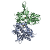
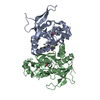
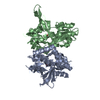
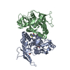
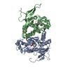
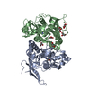
- PDB-6uz6: Crystal structure of GLUN1/GLUN2A-4M mutant ligand-binding domain... -
+ Open data

- Basic information
| Entry | Database: PDB / ID: 6uz6 | |||||||||
|---|---|---|---|---|---|---|---|---|---|---|
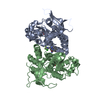
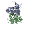
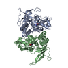
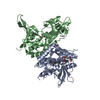
| Title | Crystal structure of GLUN1/GLUN2A-4M mutant ligand-binding domain in complex with glycine and glutamate | |||||||||
 Components Components |
| |||||||||
 Keywords Keywords | MEMBRANE PROTEIN / NMDAR / ligand-binding domain / agonist | |||||||||
| Function / homology |  Function and homology information Function and homology informationregulation of response to alcohol / response to ammonium ion / neurotransmitter receptor transport, plasma membrane to endosome / receptor recycling / response to environmental enrichment / directional locomotion / pons maturation / EPHB-mediated forward signaling / Assembly and cell surface presentation of NMDA receptors / auditory behavior ...regulation of response to alcohol / response to ammonium ion / neurotransmitter receptor transport, plasma membrane to endosome / receptor recycling / response to environmental enrichment / directional locomotion / pons maturation / EPHB-mediated forward signaling / Assembly and cell surface presentation of NMDA receptors / auditory behavior / positive regulation of Schwann cell migration / regulation of cell communication / cellular response to magnesium ion / olfactory learning / response to other organism / response to hydrogen sulfide / serotonin metabolic process / dendritic branch / conditioned taste aversion / response to methylmercury / protein localization to postsynaptic membrane / regulation of ARF protein signal transduction / response to manganese ion / transmitter-gated monoatomic ion channel activity / suckling behavior / sleep / regulation of NMDA receptor activity / response to carbohydrate / regulation of respiratory gaseous exchange / cellular response to lipid / propylene metabolic process / response to glycine / dendritic spine organization / locomotion / cellular response to dsRNA / RAF/MAP kinase cascade / positive regulation of inhibitory postsynaptic potential / response to amine / neurotransmitter receptor complex / Synaptic adhesion-like molecules / response to glycoside / regulation of monoatomic cation transmembrane transport / NMDA glutamate receptor activity / NMDA selective glutamate receptor complex / glutamate binding / voltage-gated monoatomic cation channel activity / glutamate receptor signaling pathway / ligand-gated sodium channel activity / neuromuscular process / regulation of axonogenesis / calcium ion transmembrane import into cytosol / regulation of dendrite morphogenesis / male mating behavior / regulation of synapse assembly / protein heterotetramerization / response to morphine / spinal cord development / glycine binding / startle response / dopamine metabolic process / cellular response to zinc ion / positive regulation of reactive oxygen species biosynthetic process / response to lithium ion / parallel fiber to Purkinje cell synapse / monoatomic ion channel complex / regulation of postsynaptic membrane potential / monoatomic cation transmembrane transport / positive regulation of calcium ion transport into cytosol / modulation of excitatory postsynaptic potential / cellular response to glycine / associative learning / positive regulation of dendritic spine maintenance / response to light stimulus / action potential / Unblocking of NMDA receptors, glutamate binding and activation / positive regulation of protein targeting to membrane / regulation of neuronal synaptic plasticity / monoatomic cation transport / glutamate receptor binding / ligand-gated monoatomic ion channel activity / social behavior / multicellular organismal response to stress / neuron development / conditioned place preference / phosphatase binding / long-term memory / prepulse inhibition / postsynaptic density, intracellular component / monoatomic cation channel activity / synaptic cleft / response to fungicide / calcium ion homeostasis / cellular response to manganese ion / glutamate-gated receptor activity / positive regulation of synaptic transmission, glutamatergic / glutamate-gated calcium ion channel activity / presynaptic active zone membrane / cell adhesion molecule binding / neurogenesis / excitatory synapse Similarity search - Function | |||||||||
| Biological species |  | |||||||||
| Method |  X-RAY DIFFRACTION / SYNCHROTRON / MOLECULAR REPLACEMENT / Resolution: 1.66 Å X-RAY DIFFRACTION / SYNCHROTRON / MOLECULAR REPLACEMENT / Resolution: 1.66 Å | |||||||||
 Authors Authors | Wang, J.X. / Furukawa, H. | |||||||||
| Funding support |  United States, 2items United States, 2items
| |||||||||
 Citation Citation | Journal: Nat Commun / Year: 2020 Title: Structural basis of subtype-selective competitive antagonism for GluN2C/2D-containing NMDA receptors. Authors: Wang, J.X. / Irvine, M.W. / Burnell, E.S. / Sapkota, K. / Thatcher, R.J. / Li, M. / Simorowski, N. / Volianskis, A. / Collingridge, G.L. / Monaghan, D.T. / Jane, D.E. / Furukawa, H. | |||||||||
| History |
|
- Structure visualization
| Structure viewer | Molecule: MolmilJmol/JSmol |
|---|
- Downloads & links
-Download
| PDBx/mmCIF format | 6uz6.cif.gz | 320.6 KB | Display | PDBx/mmCIF format |
|---|---|---|---|---|
| PDB format | pdb6uz6.ent.gz | 215.6 KB | Display | PDB format |
| PDBx/mmJSON format | 6uz6.json.gz | Tree view | PDBx/mmJSON format | |
| Others |  Other downloads Other downloads |
-Validation report
| Arichive directory | https://data.pdbj.org/pub/pdb/validation_reports/uz/6uz6ftp://data.pdbj.org/pub/pdb/validation_reports/uz/6uz6 | HTTPS FTP |
|---|
-Related structure data
| Related structure data |  6uzgC  6uzrC  6uzwC  6uzxC  4nf8S S: Starting model for refinement C: citing same article ( |
|---|---|
| Similar structure data |
-Links
 PDBj
PDBj


- Assembly
| Deposited unit | 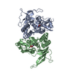
| ||||||||||||
|---|---|---|---|---|---|---|---|---|---|---|---|---|---|
| 1 |
| ||||||||||||
| Unit cell |
|
-Components
| #1: Protein | Mass: 33340.031 Da / Num. of mol.: 1 Fragment: ligand-binding domain (UNP residues 415-565,684-821) Source method: isolated from a genetically manipulated source Source: (gene. exp.)  |
|---|---|
| #2: Protein | Mass: 31606.236 Da / Num. of mol.: 1 Fragment: ligand-binding domain (UNP residues 402-539,661-800) Mutation: A414R, K738M, G740R, R741K Source method: isolated from a genetically manipulated source Source: (gene. exp.) |
| #3: Chemical | ChemComp-GLY /   Type: peptide linking / Mass: 75.067 Da / Num. of mol.: 1 / Source method: isolated from a natural source / Formula: C2H5NO2 / Feature type: SUBJECT OF INVESTIGATION Type: peptide linking / Mass: 75.067 Da / Num. of mol.: 1 / Source method: isolated from a natural source / Formula: C2H5NO2 / Feature type: SUBJECT OF INVESTIGATION |
| #4: Chemical | ChemComp-GLU /   Type: L-peptide linking / Mass: 147.129 Da / Num. of mol.: 1 / Source method: obtained synthetically / Formula: C5H9NO4 / Feature type: SUBJECT OF INVESTIGATION Type: L-peptide linking / Mass: 147.129 Da / Num. of mol.: 1 / Source method: obtained synthetically / Formula: C5H9NO4 / Feature type: SUBJECT OF INVESTIGATION |
| #5: Water | ChemComp-HOH /  Mass: 18.015 Da / Num. of mol.: 607 / Source method: isolated from a natural source / Formula: H2O Mass: 18.015 Da / Num. of mol.: 607 / Source method: isolated from a natural source / Formula: H2O |
| Has ligand of interest | Y |
| Has protein modification | Y |
-Experimental details
-Experiment
| Experiment | Method: X-RAY DIFFRACTION / Number of used crystals: 1 |
|---|
- Sample preparation
| Crystal | Density Matthews: 2.37 Å3/Da / Density % sol: 48.01 % / Description: long rod-shaped crystals |
|---|---|
| Crystal grow | Temperature: 291 K / Method: vapor diffusion, hanging drop / pH: 7 Details: 100 mM HEPES, pH 7.0, 75 mM sodium chloride, 18% PEG2000 MME |
-Data collection
| Diffraction | Mean temperature: 100 K / Serial crystal experiment: N |
|---|---|
| Diffraction source | Source: SYNCHROTRON / Site: NSLS-II / Beamline: 17-ID-1 / Wavelength: 0.92013 Å |
| Detector | Type: DECTRIS EIGER X 9M / Detector: PIXEL / Date: Apr 18, 2018 |
| Radiation | Monochromator: Si(111) / Protocol: SINGLE WAVELENGTH / Monochromatic (M) / Laue (L): M / Scattering type: x-ray |
| Radiation wavelength | Wavelength: 0.92013 Å / Relative weight: 1 |
| Reflection | Resolution: 1.66→73.1 Å / Num. obs: 71406 / % possible obs: 96.4 % / Redundancy: 6.3 % / Biso Wilson estimate: 22.21 Å2 / Rmerge(I) obs: 0.073 / Net I/σ(I): 14.3 |
| Reflection shell | Resolution: 1.66→1.69 Å / Redundancy: 3.9 % / Num. unique obs: 5481 / CC1/2: 0.515 |
- Processing
| Software |
| |||||||||||||||||||||||||||||||||||||||||||||||||||||||||||||||||||||||||||||||||||||||||||||||||||||||||||||||||||||||||||||||||||||||||||||||||||||||||||||||||||||||||||||||||||||||||||||
|---|---|---|---|---|---|---|---|---|---|---|---|---|---|---|---|---|---|---|---|---|---|---|---|---|---|---|---|---|---|---|---|---|---|---|---|---|---|---|---|---|---|---|---|---|---|---|---|---|---|---|---|---|---|---|---|---|---|---|---|---|---|---|---|---|---|---|---|---|---|---|---|---|---|---|---|---|---|---|---|---|---|---|---|---|---|---|---|---|---|---|---|---|---|---|---|---|---|---|---|---|---|---|---|---|---|---|---|---|---|---|---|---|---|---|---|---|---|---|---|---|---|---|---|---|---|---|---|---|---|---|---|---|---|---|---|---|---|---|---|---|---|---|---|---|---|---|---|---|---|---|---|---|---|---|---|---|---|---|---|---|---|---|---|---|---|---|---|---|---|---|---|---|---|---|---|---|---|---|---|---|---|---|---|---|---|---|---|---|---|---|
| Refinement | Method to determine structure: MOLECULAR REPLACEMENT Starting model: PDB entry 4NF8 Resolution: 1.66→50.07 Å / SU ML: 0.1746 / Cross valid method: FREE R-VALUE / σ(F): 1.36 / Phase error: 21.333
| |||||||||||||||||||||||||||||||||||||||||||||||||||||||||||||||||||||||||||||||||||||||||||||||||||||||||||||||||||||||||||||||||||||||||||||||||||||||||||||||||||||||||||||||||||||||||||||
| Solvent computation | Shrinkage radii: 0.9 Å / VDW probe radii: 1.11 Å | |||||||||||||||||||||||||||||||||||||||||||||||||||||||||||||||||||||||||||||||||||||||||||||||||||||||||||||||||||||||||||||||||||||||||||||||||||||||||||||||||||||||||||||||||||||||||||||
| Displacement parameters | Biso mean: 30.05 Å2 | |||||||||||||||||||||||||||||||||||||||||||||||||||||||||||||||||||||||||||||||||||||||||||||||||||||||||||||||||||||||||||||||||||||||||||||||||||||||||||||||||||||||||||||||||||||||||||||
| Refinement step | Cycle: LAST / Resolution: 1.66→50.07 Å
| |||||||||||||||||||||||||||||||||||||||||||||||||||||||||||||||||||||||||||||||||||||||||||||||||||||||||||||||||||||||||||||||||||||||||||||||||||||||||||||||||||||||||||||||||||||||||||||
| Refine LS restraints |
| |||||||||||||||||||||||||||||||||||||||||||||||||||||||||||||||||||||||||||||||||||||||||||||||||||||||||||||||||||||||||||||||||||||||||||||||||||||||||||||||||||||||||||||||||||||||||||||
| LS refinement shell |
| |||||||||||||||||||||||||||||||||||||||||||||||||||||||||||||||||||||||||||||||||||||||||||||||||||||||||||||||||||||||||||||||||||||||||||||||||||||||||||||||||||||||||||||||||||||||||||||
| Refinement TLS params. | Method: refined / Origin x: 40.641021733 Å / Origin y: 75.7939187105 Å / Origin z: 25.8618815501 Å
| |||||||||||||||||||||||||||||||||||||||||||||||||||||||||||||||||||||||||||||||||||||||||||||||||||||||||||||||||||||||||||||||||||||||||||||||||||||||||||||||||||||||||||||||||||||||||||||
| Refinement TLS group | Selection details: all |
