 Movie
Movie Controller

[English] 日本語
 Yorodumi
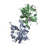
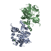
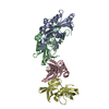
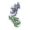
- PDB-6uw5: The crystal structure of FbiA from Mycobacterium smegmatis, GDP a... -
+ Open data

- Basic information
| Entry | Database: PDB / ID: 6uw5 | ||||||||||||
|---|---|---|---|---|---|---|---|---|---|---|---|---|---|
| Title | The crystal structure of FbiA from Mycobacterium smegmatis, GDP and Fo bound form | ||||||||||||
 Components Components | Phosphoenolpyruvate transferase | ||||||||||||
 Keywords Keywords | BIOSYNTHETIC PROTEIN / Factor 420 / Phosphotransferase / Metalloenzyme | ||||||||||||
| Function / homology |  Function and homology information Function and homology information2-phospho-L-lactate transferase / LPPG:FO 2-phospho-L-lactate transferase activity / magnesium ion binding Similarity search - Function | ||||||||||||
| Biological species |  Mycolicibacterium smegmatis (bacteria) Mycolicibacterium smegmatis (bacteria) | ||||||||||||
| Method |  X-RAY DIFFRACTION / SYNCHROTRON / MOLECULAR REPLACEMENT / Resolution: 2.2 Å X-RAY DIFFRACTION / SYNCHROTRON / MOLECULAR REPLACEMENT / Resolution: 2.2 Å | ||||||||||||
 Authors Authors | Grinter, R. / Gillett, D. / Cordero, P.R.F. / Greening, C. | ||||||||||||
| Funding support |  Australia, 3items Australia, 3items
| ||||||||||||
 Citation Citation | Journal: mSystems / Year: 2020 Title: Cellular and Structural Basis of Synthesis of the Unique Intermediate Dehydro-F420-0 in Mycobacteria. Authors: Grinter, R. / Ney, B. / Brammananth, R. / Barlow, C.K. / Cordero, P.R.F. / Gillett, D.L. / Izore, T. / Cryle, M.J. / Harold, L.K. / Cook, G.M. / Taiaroa, G. / Williamson, D.A. / Warden, A.C. ...Authors: Grinter, R. / Ney, B. / Brammananth, R. / Barlow, C.K. / Cordero, P.R.F. / Gillett, D.L. / Izore, T. / Cryle, M.J. / Harold, L.K. / Cook, G.M. / Taiaroa, G. / Williamson, D.A. / Warden, A.C. / Oakeshott, J.G. / Taylor, M.C. / Crellin, P.K. / Jackson, C.J. / Schittenhelm, R.B. / Coppel, R.L. / Greening, C. | ||||||||||||
| History |
|
- Structure visualization
| Structure viewer | Molecule: MolmilJmol/JSmol |
|---|
- Downloads & links
-Download
| PDBx/mmCIF format | 6uw5.cif.gz | 266.2 KB | Display | PDBx/mmCIF format |
|---|---|---|---|---|
| PDB format | pdb6uw5.ent.gz | 210.2 KB | Display | PDB format |
| PDBx/mmJSON format | 6uw5.json.gz | Tree view | PDBx/mmJSON format | |
| Others |  Other downloads Other downloads |
-Validation report
| Arichive directory | https://data.pdbj.org/pub/pdb/validation_reports/uw/6uw5ftp://data.pdbj.org/pub/pdb/validation_reports/uw/6uw5 | HTTPS FTP |
|---|
-Related structure data
| Related structure data |  6uvxC  6uw1C 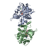 6uw3C 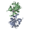 6uw7C  3c3dS C: citing same article ( S: Starting model for refinement |
|---|---|
| Similar structure data |


-Links
 PDBj
PDBj- Assembly
| Deposited unit | 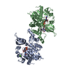
| ||||||||||||||||||||||||||||||||||||||||||||||||||||||||||||||||
|---|---|---|---|---|---|---|---|---|---|---|---|---|---|---|---|---|---|---|---|---|---|---|---|---|---|---|---|---|---|---|---|---|---|---|---|---|---|---|---|---|---|---|---|---|---|---|---|---|---|---|---|---|---|---|---|---|---|---|---|---|---|---|---|---|---|
| 1 |
| ||||||||||||||||||||||||||||||||||||||||||||||||||||||||||||||||
| Unit cell |
| ||||||||||||||||||||||||||||||||||||||||||||||||||||||||||||||||
| Noncrystallographic symmetry (NCS) | NCS domain:
NCS domain segments: Ens-ID: 1
|
-Components
| #1: Protein | Mass: 34790.613 Da / Num. of mol.: 2 Source method: isolated from a genetically manipulated source Source: (gene. exp.) Mycolicibacterium smegmatis (strain ATCC 700084 / mc(2)155) (bacteria)Strain: ATCC 700084 / mc(2)155 / Gene: fbiA, MSMEG_1830, MSMEI_1787 Production host: Mycolicibacterium smegmatis MC2 155 (bacteria)References: UniProt: A0QTG2, 2-phospho-L-lactate transferase #2: Chemical |   Type: RNA linking / Mass: 443.201 Da / Num. of mol.: 2 / Source method: obtained synthetically / Formula: C10H15N5O11P2 / Feature type: SUBJECT OF INVESTIGATION / Comment: GDP, energy-carrying molecule*YM Type: RNA linking / Mass: 443.201 Da / Num. of mol.: 2 / Source method: obtained synthetically / Formula: C10H15N5O11P2 / Feature type: SUBJECT OF INVESTIGATION / Comment: GDP, energy-carrying molecule*YM#3: Chemical | ChemComp-FO1 / | 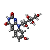  Mass: 363.322 Da / Num. of mol.: 1 / Source method: obtained synthetically / Formula: C16H17N3O7 / Feature type: SUBJECT OF INVESTIGATION Mass: 363.322 Da / Num. of mol.: 1 / Source method: obtained synthetically / Formula: C16H17N3O7 / Feature type: SUBJECT OF INVESTIGATION#4: Chemical | ChemComp-CA /   Mass: 40.078 Da / Num. of mol.: 4 / Source method: obtained synthetically / Formula: Ca / Feature type: SUBJECT OF INVESTIGATION Mass: 40.078 Da / Num. of mol.: 4 / Source method: obtained synthetically / Formula: Ca / Feature type: SUBJECT OF INVESTIGATION#5: Water | ChemComp-HOH / |  Mass: 18.015 Da / Num. of mol.: 107 / Source method: isolated from a natural source / Formula: H2O Mass: 18.015 Da / Num. of mol.: 107 / Source method: isolated from a natural source / Formula: H2OHas ligand of interest | Y | |
|---|
-Experimental details
-Experiment
| Experiment | Method: X-RAY DIFFRACTION / Number of used crystals: 1 |
|---|
- Sample preparation
| Crystal | Density Matthews: 2.21 Å3/Da / Density % sol: 44.43 % |
|---|---|
| Crystal grow | Temperature: 298 K / Method: vapor diffusion, sitting drop / Details: 18 % PEG6000, 0.1 M Tris, 0.2 M calcium acetate |
-Data collection
| Diffraction | Mean temperature: 100 K / Serial crystal experiment: N |
|---|---|
| Diffraction source | Source: SYNCHROTRON / Site: Australian Synchrotron / Beamline: MX2 / Wavelength: 0.987 Å |
| Detector | Type: DECTRIS EIGER X 16M / Detector: PIXEL / Date: Oct 29, 2019 / Details: Silicon |
| Radiation | Protocol: SINGLE WAVELENGTH / Monochromatic (M) / Laue (L): M / Scattering type: x-ray |
| Radiation wavelength | Wavelength: 0.987 Å / Relative weight: 1 |
| Reflection | Resolution: 2.2→45.864 Å / Num. obs: 30975 / % possible obs: 100 % / Redundancy: 3.5 % / CC1/2: 0.971 / Rmerge(I) obs: 0.229 / Rpim(I) all: 0.168 / Net I/σ(I): 3.1 |
| Reflection shell | Resolution: 2.2→2.27 Å / Redundancy: 3.5 % / Rmerge(I) obs: 2.204 / Mean I/σ(I) obs: 0.7 / Num. unique obs: 2644 / CC1/2: 0.175 / Rpim(I) all: 1.623 / % possible all: 100 |
- Processing
| Software |
| ||||||||||||||||||||||||||||||||||||||||||||||||||||||||||||||||||||||||
|---|---|---|---|---|---|---|---|---|---|---|---|---|---|---|---|---|---|---|---|---|---|---|---|---|---|---|---|---|---|---|---|---|---|---|---|---|---|---|---|---|---|---|---|---|---|---|---|---|---|---|---|---|---|---|---|---|---|---|---|---|---|---|---|---|---|---|---|---|---|---|---|---|---|
| Refinement | Method to determine structure: MOLECULAR REPLACEMENT Starting model: 3C3D Resolution: 2.2→45.821 Å / SU ML: 0.32 / Cross valid method: THROUGHOUT / σ(F): 1.33 / Phase error: 28.6 / Stereochemistry target values: ML
| ||||||||||||||||||||||||||||||||||||||||||||||||||||||||||||||||||||||||
| Solvent computation | Shrinkage radii: 0.9 Å / VDW probe radii: 1.11 Å / Solvent model: FLAT BULK SOLVENT MODEL | ||||||||||||||||||||||||||||||||||||||||||||||||||||||||||||||||||||||||
| Displacement parameters | Biso max: 93.98 Å2 / Biso mean: 41.3258 Å2 / Biso min: 22.32 Å2 | ||||||||||||||||||||||||||||||||||||||||||||||||||||||||||||||||||||||||
| Refinement step | Cycle: final / Resolution: 2.2→45.821 Å
| ||||||||||||||||||||||||||||||||||||||||||||||||||||||||||||||||||||||||
| Refine LS restraints NCS |
| ||||||||||||||||||||||||||||||||||||||||||||||||||||||||||||||||||||||||
| LS refinement shell | Refine-ID: X-RAY DIFFRACTION / Rfactor Rfree error: 0
| ||||||||||||||||||||||||||||||||||||||||||||||||||||||||||||||||||||||||
| Refinement TLS params. | Method: refined / Origin x: 39.0302 Å / Origin y: 14.2258 Å / Origin z: 34.4565 Å
| ||||||||||||||||||||||||||||||||||||||||||||||||||||||||||||||||||||||||
| Refinement TLS group |
|
