 Movie
Movie Controller

[English] 日本語
 Yorodumi
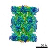
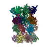
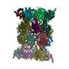
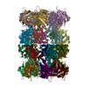
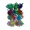
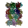
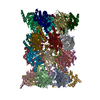
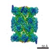
- PDB-6uti: Allosteric coupling between alpha-rings of 20S proteasome, 20S pr... -
+ Open data

- Basic information
| Entry | Database: PDB / ID: 6uti | ||||||||||||||||||||||||||||||||||||||||||||||||||||||||||||
|---|---|---|---|---|---|---|---|---|---|---|---|---|---|---|---|---|---|---|---|---|---|---|---|---|---|---|---|---|---|---|---|---|---|---|---|---|---|---|---|---|---|---|---|---|---|---|---|---|---|---|---|---|---|---|---|---|---|---|---|---|---|
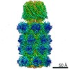
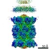
| Title | Allosteric coupling between alpha-rings of 20S proteasome, 20S proteasome with singly capped PAN complex | ||||||||||||||||||||||||||||||||||||||||||||||||||||||||||||
 Components Components |
| ||||||||||||||||||||||||||||||||||||||||||||||||||||||||||||
 Keywords Keywords | HYDROLASE / PAN / proteasome / singly-capped | ||||||||||||||||||||||||||||||||||||||||||||||||||||||||||||
| Function / homology |  Function and homology information Function and homology informationproteasome endopeptidase complex / proteasome core complex, beta-subunit complex / threonine-type endopeptidase activity / proteasome core complex, alpha-subunit complex / proteasomal protein catabolic process / endopeptidase activity / ubiquitin-dependent protein catabolic process / cytoplasm Similarity search - Function | ||||||||||||||||||||||||||||||||||||||||||||||||||||||||||||
| Biological species |   Thermoplasma acidophilum (acidophilic) Thermoplasma acidophilum (acidophilic) | ||||||||||||||||||||||||||||||||||||||||||||||||||||||||||||
| Method | ELECTRON MICROSCOPY / single particle reconstruction / cryo EM / Resolution: 3.4 Å | ||||||||||||||||||||||||||||||||||||||||||||||||||||||||||||
 Authors Authors | Cheng, Y. / Yu, Z. | ||||||||||||||||||||||||||||||||||||||||||||||||||||||||||||
| Funding support |  United States, 1items United States, 1items
| ||||||||||||||||||||||||||||||||||||||||||||||||||||||||||||
 Citation Citation | Journal: Nat Commun / Year: 2020 Title: Allosteric coupling between α-rings of the 20S proteasome. Authors: Zanlin Yu / Yadong Yu / Feng Wang / Alexander G Myasnikov / Philip Coffino / Yifan Cheng / Abstract: Proteasomal machinery performs essential regulated protein degradation in eukaryotes. Classic proteasomes are symmetric, with a regulatory ATPase docked at each end of the cylindrical 20S. Asymmetric ...Proteasomal machinery performs essential regulated protein degradation in eukaryotes. Classic proteasomes are symmetric, with a regulatory ATPase docked at each end of the cylindrical 20S. Asymmetric complexes are also present in cells, either with a single ATPase or with an ATPase and non-ATPase at two opposite ends. The mechanism that populates these different proteasomal complexes is unknown. Using archaea homologs, we construct asymmetric forms of proteasomes. We demonstrate that the gate conformation of the two opposite ends of 20S are coupled: binding one ATPase opens a gate locally, and also opens the opposite gate allosterically. Such allosteric coupling leads to cooperative binding of proteasomal ATPases to 20S and promotes formation of proteasomes symmetrically configured with two identical ATPases. It may also promote formation of asymmetric complexes with an ATPase and a non-ATPase at opposite ends. We propose that in eukaryotes a similar mechanism regulates the composition of the proteasomal population. | ||||||||||||||||||||||||||||||||||||||||||||||||||||||||||||
| History |
|
- Structure visualization
| Movie |
Movie viewer |
|---|---|
| Structure viewer | Molecule: MolmilJmol/JSmol |
- Downloads & links
-Download
| PDBx/mmCIF format | 6uti.cif.gz | 968.3 KB | Display | PDBx/mmCIF format |
|---|---|---|---|---|
| PDB format | pdb6uti.ent.gz | 822.1 KB | Display | PDB format |
| PDBx/mmJSON format | 6uti.json.gz | Tree view | PDBx/mmJSON format | |
| Others |  Other downloads Other downloads |
-Validation report
| Arichive directory | https://data.pdbj.org/pub/pdb/validation_reports/ut/6utiftp://data.pdbj.org/pub/pdb/validation_reports/ut/6uti | HTTPS FTP |
|---|
-Related structure data
| Related structure data | 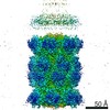 20880MC  6utfC  6utgC  6uthC  6utjC M: map data used to model this data C: citing same article ( |
|---|---|
| Similar structure data |


-Links
 PDBj
PDBj


- Assembly
| Deposited unit | 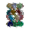
|
|---|---|
| 1 |
|
-Components
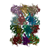
| #1: Protein | Mass: 25067.518 Da / Num. of mol.: 7 Source method: isolated from a genetically manipulated source Source: (gene. exp.) Thermoplasma acidophilum (acidophilic) / Gene: psmA, Ta1288 / Production host:  Escherichia phage EcSzw-2 (virus) Escherichia phage EcSzw-2 (virus)References: UniProt: P25156, proteasome endopeptidase complex #2: Protein | Mass: 25081.584 Da / Num. of mol.: 7 / Mutation: R28L Source method: isolated from a genetically manipulated source Source: (gene. exp.) Thermoplasma acidophilum (acidophilic) / Gene: psmA, Ta1288 / Production host: Escherichia phage EcSzw-2 (virus)References: UniProt: P25156, proteasome endopeptidase complex #3: Protein | Mass: 22294.848 Da / Num. of mol.: 14 Source method: isolated from a genetically manipulated source Source: (gene. exp.) Thermoplasma acidophilum (acidophilic) / Gene: psmB, Ta0612 / Production host: Escherichia phage EcSzw-2 (virus)References: UniProt: P28061, proteasome endopeptidase complex Has protein modification | N | |
|---|
-Experimental details
-Experiment
| Experiment | Method: ELECTRON MICROSCOPY |
|---|---|
| EM experiment | Aggregation state: PARTICLE / 3D reconstruction method: single particle reconstruction |
- Sample preparation
| Component | Name: Complex of 20S proteasome with singly capped PAN / Type: COMPLEX / Entity ID: all / Source: RECOMBINANT |
|---|---|
| Source (natural) | Organism: Thermoplasma acidophilum (acidophilic) |
| Source (recombinant) | Organism: Escherichia phage EcSzw-2 (virus) |
| Buffer solution | pH: 7.5 |
| Specimen | Embedding applied: NO / Shadowing applied: NO / Staining applied: NO / Vitrification applied: YES |
| Specimen support | Details: unspecified |
| Vitrification | Cryogen name: ETHANE |
- Electron microscopy imaging
| Experimental equipment |  Model: Tecnai Polara / Image courtesy: FEI Company |
|---|---|
| Microscopy | Model: FEI POLARA 300 |
| Electron gun | Electron source:  FIELD EMISSION GUN / Accelerating voltage: 300 kV / Illumination mode: FLOOD BEAM FIELD EMISSION GUN / Accelerating voltage: 300 kV / Illumination mode: FLOOD BEAM |
| Electron lens | Mode: BRIGHT FIELD |
| Image recording | Electron dose: 60 e/Å2 / Film or detector model: GATAN K2 SUMMIT (4k x 4k) |
- Processing
| Software | Name: PHENIX / Version: 1.12_2829: / Classification: refinement | ||||||||||||||||||||||||
|---|---|---|---|---|---|---|---|---|---|---|---|---|---|---|---|---|---|---|---|---|---|---|---|---|---|
| EM software | Name: PHENIX / Category: model refinement | ||||||||||||||||||||||||
| CTF correction | Type: NONE | ||||||||||||||||||||||||
| 3D reconstruction | Resolution: 3.4 Å / Resolution method: FSC 0.143 CUT-OFF / Num. of particles: 33000 / Symmetry type: POINT | ||||||||||||||||||||||||
| Refine LS restraints |
|
